2D/3D probing of hierarchically structured functional materials using complementary X-ray and electron microscopy
- Abstract number
- 1385
- Event
- European Microscopy Congress 2020
- DOI
- 10.22443/rms.emc2020.1385
- Corresponding Email
- [email protected]
- Session
- PST.6 - In-situ and in-operando microscopy
- Authors
- Yakub Fam (4, 2), Thomas Sheppard (2), Jan-Dierk Grunwaldt (2), Andreas Schropp (1), Christian G. Schroer (1), Mirko Holler (3), Ana Diaz (3)
- Affiliations
-
1. Deutsches Elektronen-Synchrotron
2. Karlsruhe Institute of Technology
3. Paul Scherrer Institut
4. Technical University of Denmark
- Keywords
In-situ, tomography, X-ray ptychography, electron microscopy, quantitative analysis, simulation
- Abstract text
Functional materials, such as catalysts, are commonly materials with hierarchical structures, so characterizations at various length scales and especially under working conditions are important to improve their performances as well as to reveal their working mechanisms [1]. Moreover, combining several techniques from different sources to obtain complementary information is necessary [2, 3] due to the unavailability of one technique capable of covering all the length scales. In the case of length/size characterization techniques, microscopy has been considered to be the best means to investigate functional materials either in 2D or more preferably in 3D owing to the capability to simultaneously probe their morphologies, electronic spectra, and even diffraction patterns.
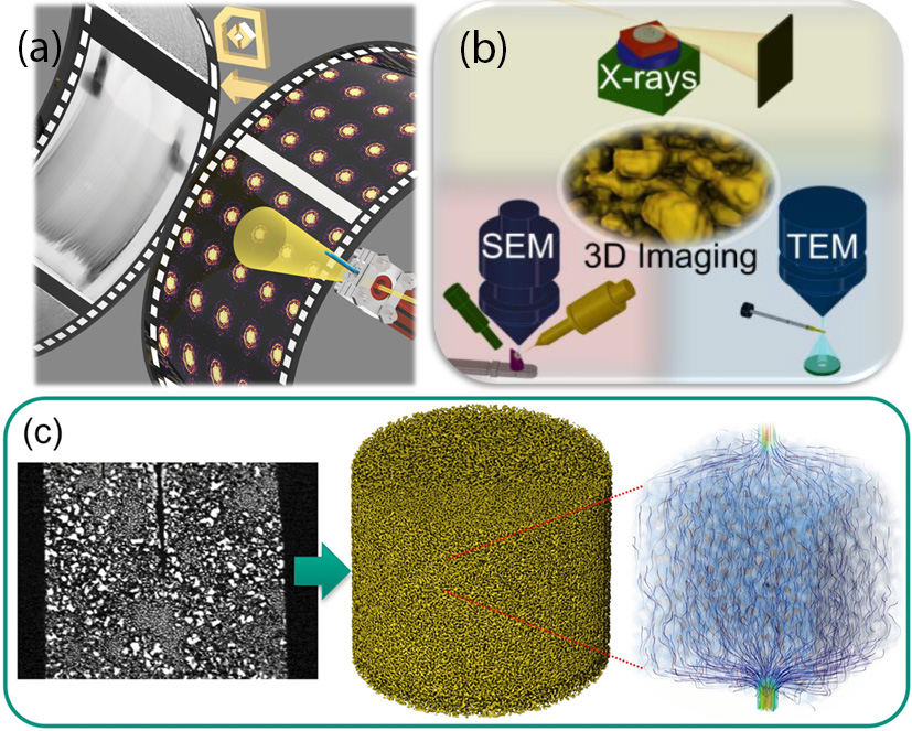
The work started with exploring three different ex situ 3D imaging techniques, i.e. ptychographic X-ray computed tomography (PXCT), electron tomography (ET), and focused ion beam slice-and view (FIB-SV). It utilized nanoporous gold as the model and it unravelled the versatility of 3D microscopy beyond imaging purpose with each technique has its own pros and cons. This first part also pointed out the importance of complementary nature between electron and X-ray microscopy, where electron results in higher spatial resolution and X-ray encompasses larger field of view. The second part focused on PXCT for quasi in situ thermal treatment of the same materials at atmospheric condition. It highlighted its ease of use for future in situ implementation, also complemented by ET to strengthen its advantage in resolving what PXCT is not able to. The 3D rendered data were then used for fluid flow and thermal conductivity simulations to further reveal another benefit from 3D imaging. The last part of the work brings about the development of in situ cell to realize the complete in situ studies under controlled temperatures and environments. The cell was built for its capability to conduct both 2D X-ray ptychography (XRP) experiments and limited-angle PXCT with simultaneous X-ray fluorescence mapping. The feasibility tests made use of functional materials, i.e. nanoporous gold, zeolite, and cobalt-manganese-oxides hollow sphere, as the models for thermal annealing treatment under certain atmospheres. This last work ultimately verifies the necessity of in situ studies to visualize the morphology online either in 2D or 3D, to also map the elemental distributions live, and more importantly to accurately determining the onset annealing temperatures under specific atmospheres. Furthermore, transmission electron microscopy (TEM) was also performed on the same sample as a complementary technique, which brings another benefit in using the in situ cell. Regarding the in situ limited angle 3D PXCT experiment, a machine-learning tomographic reconstruction technique [5] has been developed by DESY scientists as an excellent solution to reconstruct 3D tomograms with shortened acquisition times despite the lack of projection images at higher tilting angles.
Figure 1. (a) Illustration of in situ cell for in situ X-ray ptychography experiment with heating and gas flow treatment at P06 beamline (DESY) with its resulting diffraction patterns, then turned into ptychograms of nanoporous gold [4]; (b) Illustration of complementary electron (FIB-SV and ET) and X-ray microscopy (PXCT) on nanoporous gold to reveal both morphology and physical properties in 3D [2]; (c) Tomogram, 3D rendering, fluid-flow simulation profile of nanoporous gold from PXCT measurement at cSAXS beamline (PSI).
To conclude, this work converges into the ideal study of conducting all-around in situ 3D imaging of functional materials for quantitative analysis and simulation. The future realization of using the in situ cell will be operando 3D complementary X-ray and electron microscopic studies of functional materials, for example porous zeolites for CO2 capture and conversion.
- References
[1] J.-D. Grunwaldt et al, "Imaging Catalysts at Work: A Hierarchical Approach from the Macro- to the Meso- and Nano-scale" ChemCatChem 5 (2013) 62-80.
[2] Y. Fam et al, "Correlative Multiscale 3D Imaging of a Hierarchical Nanoporous Gold Catalyst by Electron, Ion and X-ray Nanotomography" ChemCatChem 10 (2018) 2858-2867.
[3] T. Li et al, "Composition and Structure Dependent Mesopore/Macropore Formation in Zeolites by Desilication" J. Phys. Chem. C 123 (2019) 8793-8801.
[4] Y. Fam et al, "A versatile nanoreactor for complementary in situ X-ray and electron microscopy studies in catalysis and materials science" J. Synchrotron Radiat. 26 (2019) 1769-1781.
[5] X. Yang et al, “Tomographic reconstruction with a generative adversarial network” J. Synchrotron Radiat. 27 (2020) 486-493.
[6] The authors gratefully acknowledge funding from the German’s Federal Ministry of Education and Research, “MicTomoCat” (05K16VK1).