3D characterization of heat-induced morphological changes of Au nanostars by fast in-situ electron tomography
- Abstract number
- 1294
- Event
- Virtual Early Career European Microscopy Congress 2020
- Presentation Form
- Submitted Oral
- DOI
- 10.22443/rms.emc2020.1294
- Corresponding Email
- [email protected]
- Session
- PST.6 - In-situ and in-operando microscopy
- Authors
- Hans Vanrompay (3, 5), Dr. Eva Bladt (3, 5), Dr. Wiebke Albrecht (3, 5), Dr. Armand Béché (3, 5), Dr. Marina Zakhozheva (2), Dr. Ana Sánchez-Iglesias (1), Prof. Dr. Luis M. Liz-Marzán (1, 4), Prof. Dr. Sara Bals (3, 5)
- Affiliations
-
1. CIC biomaGUNE and Ciber-BBN, Basque Research and Technology Alliance (BRTA)
2. DENSsolutions
3. Electron Microscopy for Materials Science (EMAT), University of Antwerp
4. Ikerbasque, Basque Foundation for Science
5. NANOlab Center of Excellence, University of Antwerp
- Keywords
In-situ heating, HAADF-STEM tomography, Au nanostars, plasmonics
- Abstract text
Introduction
Gold nanoparticles (Au NPs) have shown enormous potential for applications in various fields, ranging from biology and medicine to chemistry and physics. Next to their catalytic activity, high chemical stability and bio-compatibility, Au NPs exhibit intriguing optical properties due to well-defined localized surface plasmon resonances (LSPRs). The plasmonic properties of Au NPs can be tuned by varying their morphology. One exciting example of plasmonic NPs are anisotropic NPs such as nanostars (NSs). Due to their morphology they exhibit superior electromagnetic field enhancements, but are known to be unstable during thermal or photothermal treatment needed in e.g. cancer treatment or catalysis. Such heat-induced deformations will consequently affect the optical properties of the Au NSs and limit their applicability. It is therefore of great importance to understand the heat-induced morphological evolution of highly anisotropic Au NPs and the correlated change of their optical properties.
Up to date, most studies have focused on understanding the (photo)thermal stability of large ensembles of Au NPs. So far, these studies have been consistently based on conventional, two-dimensional (2D) images, which inherently limit such investigations to simple geometries such as rods or spheres. To monitor the deformation of more complicated anisotropic structures such as the Au NSs, 2D projection images are insufficient.
Electron tomography is currently a standard technique to visualize the morphology and internal structure of a wide variety of nanostructures in three dimensions (3D). One remaining limitation to perform such a 3D in-situ study is that the necessary time for a standard tomographic experiment is typically too long to follow the induced morphological changes in 3D. Conventionally, electron tomography experiments are based on acquiring tilt series of projection images with an increment of 1-2˚ over an as large as possible angular range. Although the acquisition of such a tilt series is mostly automated, even under ideal conditions approximately 1 hour is required to obtain all images. This is clearly a major drawback when trying to connect the properties of the nanoparticles to their 3D structure.
Objective
We therefore propose an acquisition approach where a tilt series of 2D high angle annular dark field scanning transmission electron microscopy (HAADF-STEM) projection images is acquired within a few minutes. By continuously tilting the holder and simultaneously acquiring projection images while focusing and tracking the particle, we were able to reduce the total acquisition time for a tilt series by a factor of ten. In this manner, we were able to study the 3D morphological evolution of single Au NSs as a function of both heating time and temperature.
Materials and Methods
The electron tomography experiments were performed using an aberration-corrected cubed Thermo Fisher Scientific Titan electron microscope operated 300 kV and a DENSsolutions tomography heating holder with MEMS-based heating chips.
Results
Experiments were performed at 200 °C, 300 °C and 400 °C for a total heating time of 20 minutes, in order to decouple the effect of temperature and total amount of delivered heat on the morphological changes. Fast tilt series were acquired at intermediate time steps of 30 s, 60 s, 90 s, 120 s, 180 s, 300 s, 600 s and 1200 s for each temperature. Hence, the particle was heated for the given time and subsequently cooled down to room temperature, so as to quench further heat-induced morphological transformations. After quenching, a fast tomographic series was acquired. This procedure was repeated for the various selected time steps.
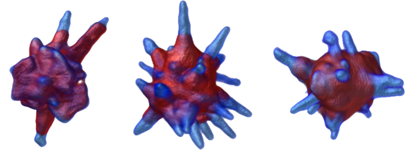
We were thereby able to quantify local volume reductions and increments, as well as the determination of the local curvatures of NSs during heating. At elevated temperatures, the sharp tips at the end of the nanobranches were observed to reshape into shorter and blunter tips. While it has been previously postulated by indirect methods that curvature-induced surface diffusion is the driving mechanism of such reshaping, our experiments directly confirm this hypothesis. We additionally showed that the major part of the morphological evolution occurs within the first minute of heating, regardless of temperature. The shape transition subsequently slows down and a stable final morphology is reached. The amount of total reshaping was found to increase for higher temperatures and the final morphology of the NSs to depend on the heating temperature, which thus has a more significant influence than the heating time
Figure 1: Illustration of the thermal reshaping behavior of gold nanostars.
Conclusion
We implemented a methodology to perform fast electron tomography in HAADF-STEM mode, which allowed us to observe the morphological evolution of an Au NS when subjected to heating. Due to the highly reduced acquisition time of the tilt series, gradual changes at distinct time steps during heating were investigated, which allowed us to record the 3D morphological changes as a function of heating time and temperature. We expect that this approach will be essential to investigate other dynamical processes under different in-situ conditions.
Acknowledgements
H.V. acknowledges financial support by the Research Foundation Flanders (FWO grant 1S32617N). E.B. acknowledges a post-doctoral grant from the Research Foundation Flanders (FWO, Belgium). W.A. acknowledges an Individual Fellowship funded by the Marie Sklodowska-Curie Actions (MSCA) in Horizon 2020. The authors acknowledge funding from European Commission Grant (EUSMI 731019). S.B. acknowledges financial support from European Research Council (ERC Consolidator Grant #815128-REALNANO).
- References
References
H. Vanrompay, E. Bladt, W. Albrecht, A. Béché, M. Zakhozheva, A. Sánchez-Iglesias, L.M. Liz-Marzán, S. Bals, Nanoscale 10 (2018) 22792–22801.