3D Electron Ptychographical Tomography
- Abstract number
- 1146
- Event
- European Microscopy Congress 2020
- DOI
- 10.22443/rms.emc2020.1146
- Corresponding Email
- [email protected]
- Session
- PST.5 - Diffraction techniques and structural analysis
- Authors
- Mr Zhiyuan Ding (1), Dr Si Gao (1), Prof Xiaoqing Pan (2), Prof Angus. I. Kirkland (3, 4, 5), Prof Peng Wang (1)
- Affiliations
-
1. Nanjing University
2. University of California
3. University of Oxford
4. The Rosalind Franklin Institute
5. Electron Physical Sciences Imaging Centre
- Keywords
Phase Imaging, Ptychography, Tomography, 4D-STEM
- Abstract text
Electron tomography has become a widely used and powerful technique for three dimensional (3D) structural analysis in both physical and biological sciences for decades. However, there are still remaining challenging in the areas, such as beam sensitive or low atomic number materials. In this work, we demonstrated that defocused-probe ptychography can produce 3D images from weak-scattering samples at a low dose, based upon optical sectioning and tilt series approaches, respectively.
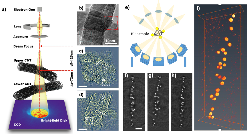
Since the development of aberration correctors, scanning transmission electron microscopy (STEM) has become a widely used and powerful technique for materials science research. Using standard high angle annular dark field STEM imaging, 3D electron tomography which recently has been demonstrated at atomic resolution provides a fundamental understanding of structure-property relationships at the atomic level. However, there are still remaining challenging in the areas such as 3D imaging of beam sensitive materials or low atomic number materials. Electron ptychography which is one of the emerged new imaging techniques has advantages of super-resolution [1,2], high phase sensitivity [3], low-dose [4]and the capability of integrating with spectroscopy[5]. In the focused-probe type of ptychography, scanning arrays of the probe are required to satisfy the sampling with a reasonable field of view, which can cause the total acquisition time and electron dose on the sample to be typically high. Therefore, it becomes ever more difficult to combine this type of ptychography further with tomography, where the recording of an entire tilt series is needed for tomographic reconstruction. Instead, using defocused-probe ptychography as shown in Fig. 1 (a), a probe defocused by a few tens of nm’s can be scanned over a large area of a specimen with highly overlapping probe positions. As a result, just a few hundreds of probe positions are sufficient to reconstruct a 2D phase. It has shown success in imaging light atoms together with heavy atoms in a LaB6 crystal [3] and MoS2 at a very low dose [4]. A new generation of fast direct electronic detectors combined with the defocused nature of the probe and few probe positions offer the potential of 3D phase imaging as well as greatly reducing the size of the collected data set.
In this work, we firstly demonstrated that defocused-probe ptychography using an inversed multi-slice method is able to produce 3D optically sectioned images of a thick sample[6], as shown in Fig. 1 (a). The experiment was carried out on a sample of carbon nanotube bundles (CNTs) as shown in Fig. 1 (b) at 60kV. Using the algorithm of 3PIE [7], the phases of the sample at different sample heights as shown in Fig. 1 (c) and d) were reconstructed. The method provides high contrast, quantitative phase maps at close to atomic lateral resolution and with a few tens of nanometers depth resolution.
Secondly, by combining ptychography with the tilt series based approach, we will show 3D phase-contrast tomography on gold particles dispersed a carbon thin film as shown in Fig. 1 (e). A tomographic tilt series of ptychographic data with a tilt range from -57o to + 52 o were collected from the particles. A tilt series of total 55 phase images were reconstructed, typically as shown in Fig. 1 (f)-(h). After being aligned, the tilt series were used to compute a 3D tomographic reconstruction using BP algorithm in IMOD[8] software and visualized, as shown in Fig. 1 (i).
Furthermore, we will implement ptychographic tomography to reconstruct the 3D structure of DNA origami assemblies with high phase sensitivity and at low doses. Therefore, we expect that electron ptychography potentially paves a way to determine the 3D structure of the weak scattering and beam-sensitive materials, such as MOF, cells and viruses, in conjunction with tilt series tomography.
Figure 1. (a) Schematic of the experimental set-up of defocused probe ptychography; (b) TEM image of the CNTs; (c) and (d) Ptychographic reconstructed phase of the upper and lower CNTs at Gaussian focus, respectively using an inversed multi-slice method. Scale bars: 10 nm. (e) Schematic of the experimental set-up of tilt series ptychographic tomography. (f)-(h) 2D Ptychographic reconstructed phase of the gold particles on a carbon thin film at the tilt angles of -57o, 0 o and 52 o, respectively. The scale bar is 20 nm. (i) 3D rendering of the tomographic reconstruction of the gold particles.
- References
[1] PD Nellist et al., Nature 374 (1995) p.630.
[2] Y Jiang et al., Nature 559 (2018) p.343.
[3] P Wang et al., Scientific Reports 7 (2017) p.2857.
[4] J Song et al., Scientific Reports 9 (2019) p.3919.
[5] B Song et al., Physical Review Letters 121 (2018) p.146101.
[6] S Gao et al., Nature Communications 8 (2017) p.163.
[7] AM Maiden et al., J. Opt. Soc. Am. A 29 (2012) p.1606.
[8] JR Kremer et al., J. struct. Bio., 116 (1996) p.0.
[9] The authors acknowledge funding from the National Natural Science Foundation of China (11874199).