3D quantification of multi-scale porosity in diatoms structures
- Abstract number
- 864
- Event
- European Microscopy Congress 2020
- DOI
- 10.22443/rms.emc2020.864
- Corresponding Email
- [email protected]
- Session
- DHA.2 - Advances in 3-dimensional image reconstruction
- Authors
- Mr Othmane Darouich (3), Mr Dris Ihiawakrim (3), Mr Walid Baaziz (3), Ms Hedwige Poncet (4), Mr Jacques Faerber (3), Ms Daniele Spehner (2), Ms Corinne Petit (1), Mr Charles Hirlimann (3), Mr Ovidiu Ersen (3)
- Affiliations
-
1. École européenne de chimie, polymères et matériaux de Strasbourg
2. Institut de Génétique et de biologie moléculaire et cellulaire
3. Institut de Physique et Chimie des Matériaux de Strasbourg
4. Institut français du pétrole
- Keywords
- Abstract text
Materials displaying a multi-scale and/or a hierarchical porosity do find several applications in fields such as biomineralization, environmental catalysis and biosensor design1. This work is devoted to designing a new characterization methodology able to properly analyze such multiscale specimens. This approach consists in the complementary use of: electron tomography, Focused Ion Beam-Scanning Electron Microscopy (FIB-SEM) and ATUMtome-based technique2 . It was firstly tested on a model unicellular organism that is coscinodiscus diatom with a structure made of periodically-ranged amorphous biosilica valve (shell). Characterized by a multiscale porosity ranging from 1 nm to several μm, these natural systems can be used as support material in various types of applications, in particular in the catalysis field3. The quantitative use of this combined analysis approach actually permitted a full characterization of the porosity of the material and led us to the discovery of an ultimate nanometric porosity that was not reported yet in the literature.
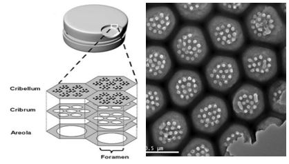
Due to their multiscale 3d structure, diatoms are able to sort and confine a large variety of particles ranging from large abundant bacteria to small molecules such as nitrates4, passing through intermediate particles in size like macromolecules or viruses5 6. In this work we focused our attention on the diatoms species called ‘coscinodiscus’. Mainly, the fractal microstructure of these diatoms does incorporate three layers into their valve structures (areolae, cribellum, cribrum) (figure 1)7. The pore size and the porosity of this species change gradually from the inner layer to the outer layer.
Figure 1. Schematic representation (left) of the three-dimensional architecture of the valve of a centric diatom, made of a honeycomb-like chambers called areolae, the hole in the floor of the areolae representing the foramen. The roof of the areolae is called the cribrum. The layer over the cribrum is called the cribellum, image taken from Losic, D et al. (2006). 2D TEM image (right) of the structure of the valve (cribrum, cribelum, areolae).
First of all, we quantified the nanoporosity using transmission electron microscopy (TEM) in the tomographic mode and the macroporosity using FIB-SEM going through the mesoporosity. Then we studied the periodicity of the pores at a larger scale by using the ATUM-SEM approach combining SEM imaging and the ATUMtome tool used here for one of the first time in the field of materials sciences.
Thereafter, we developed a comprehensive strategy allowing for the analysis and the quantification of multiscale porosities. Two types of functions were used to describe the structure of this material. The first one is based on the morphology of the individual entities while the second is used for describing its topology. Regarding the morphological parameters, we used the some classical pair distribution functions that statistically characterize a 3D microstructure. On the other hand, the topological parameters were used to describe the way the pores are connected one to the other.
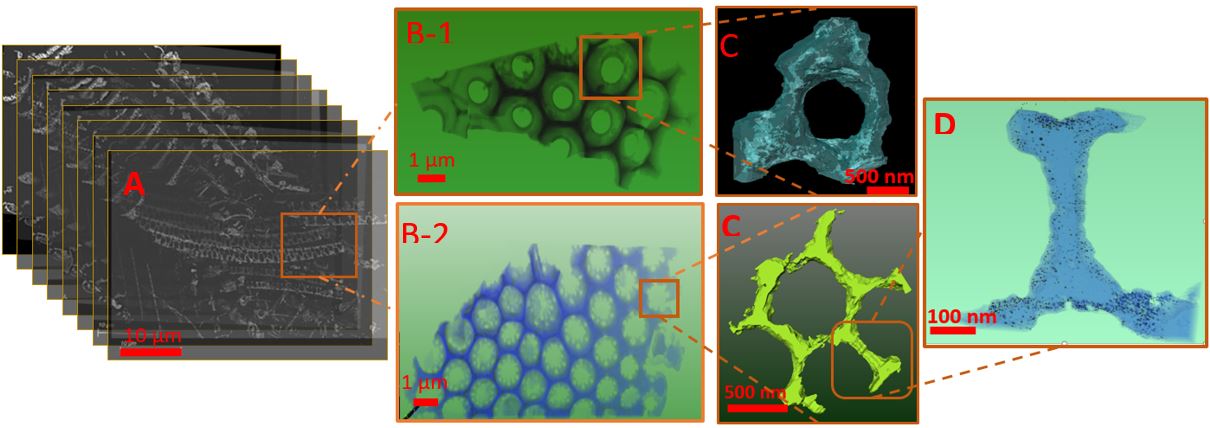
The combination of the three 3D approaches shows that this centric diatom can be referenced as a system characterized by several types of symmetry elements (figure 2). With the 3D ATUM-SEM approach, we quantified the porosity in a large part of the valve of the diatom and observed the inner layer (areolae) has a quasi-periodic honeycomb-like structure. This structure is not perfect and hence, the pore size of the areolae does suffer some dispersion around their mean values. The areolae do contain holes called foramen. The outer layer (cribrum) is made of an array of honeycomb hexagons. At this regard, we obtained more reliable information about the morphology of these two interconnected layers by using the tomographic approach of the FIB-SEM technique. It allowed us to visualize the third layer called cribellum, presenting circular pores with an average diameter of 50 nm. At a smaller scale, by using the TEM tomography, we could clearly evidence the morphology of these circular mesoporous. From a general point of view, our results clearly confirm the assumptions reported in the literature regarding the internal structures of these hierarchical systems8,9. In addition, the TEM tomography (figure D) allowed us to demonstrate that the three layers are made out of small meso-micropores (less than 7 nm in diameter). In order to study the connectivity and distribution of these small pores, quantitative studies are being carried out.
Figure 2. (A) Serial sections obtained using the ATUMtome-SEM approach, the sections do contain different species of diatoms; we focused on large part of the valve of a centric diatom. (B) 3D reconstructed FIB-SEM image of the structure of the valve, with B-1 representing the honeycomb structure associated to the cribelum and areolae and B-2 the hexagonal structures characterized by the presence of circular pores at the cribrum layer. (C) 3D reconstructed TEM view representing one areolae and hexagonal structure of the valve. (D) 3D TEM reconstruction representing a typical area of the hexagonal structure in which pores with diameters less than 7 nm can be observed.
From a materials perspective, the combination of these complementary 3D approaches gave rise to a multi-scale description of the porosity of the coscinodiscus diatom valve which is unavoidable for a clear understanding of its structure at different scales. In particular, at the nanometric scale, it was found that all the constituting layers of the diatoms do exhibit nano-pores with diameters less than 7 nm.
- References
[1] James G. Mitchell in "Diatom Nanotechnology : Progress and Emerging Applications", ed. Dusan Losic (Royal Society of Chemistry, London) p. 270.
[2] Hayworth, K. J. et al., 8 (2014), p. 68.
[3] Fischer,C. et al.,6 (2016), p. 1253-1261 .
[4] Falkowski, P. G. ; Fenchel, T. ; Delong, E. F., 320 (2008), p. 1034-1039.
[5] Hennes, K. P. ; Suttle, C. A., 6 (1995), p. 1050-1055.
[6] T. A. Sarma in "Handbook of Cyanobacteria", ed. T. A. Sarma (CRC Press).
[7] Seckbach, J. in "Diatoms: Fundamentals and Applications", ed. Seckbach, J.; Gordon, R. (Wiley, Massachusetts).
[8] Gibaud, A. et al., 17 (2019), p.2468-0230.
[9] Mcheik, A et al., 5 (2018), p.123.