A label-free multimodal nonlinear microscope for biological applications
- Abstract number
- 1337
- Event
- Virtual Early Career European Microscopy Congress 2020
- Presentation Form
- Submitted Oral
- DOI
- 10.22443/rms.emc2020.1337
- Corresponding Email
- [email protected]
- Session
- LSA.1 - Label-free life science imaging
- Authors
- Benedetta Talone (2), Valentina Parodi (1), Carlo Valensise (2), Giulio Cerullo (2), Dario Polli (2)
- Affiliations
-
1. Department of Chemistry, Materials and Chemical Engineering ''G. Natta'', Politecnico di Milano
2. Department of Physics, Politecnico di Milano
- Keywords
Coherent Raman Scattering, multimodal imaging, nonlinear microscopy
- Abstract text
INTRODUCTION: We developed a home-made, innovative and label-free transmission inverted multimodal nonlinear microscope with off-the-shelf components able to perform two-photon excitation fluorescence (TPEF), second harmonic generation (SHG), coherent anti-Stokes Raman scattering (CARS) and stimulated Raman scattering (SRS) microscopy [1]. This set-up is highly versatile, can be easily reconfigured and allows the observation of vital, complex and thick biological samples without invasive laser sources and staining. In this work, we performed nonlinear imaging of different biological samples, from single cells to highly heterogeneous tissues to validate our acquisition system.
METHODS: The microscope is fed by a multi-branch Erbium-doped amplified fiber laser. In the CARS/SRS modalities, pump (at 780nm) and tunable Stokes pulses (in the 940-1200nm range) with ps duration are employed, thus covering the CH-stretch region (2500-3200cm-1). TPEF/SHG signals are excited by a third branch delivering 100fs pulses at 780nm. The sample is mounted on an x-y translational stage, enabling us to image large sample areas.
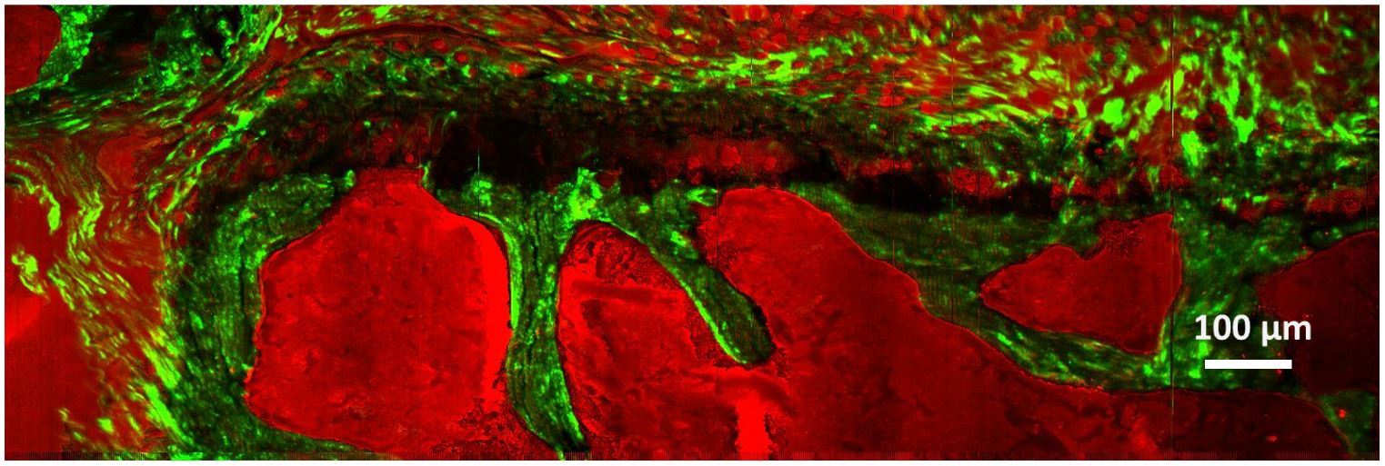
RESULTS: Figure 1 shows results on a murine vertebra section. Distribution of collagen is detected in the SHG modality (green areas), while CARS signal at 2940cm-1 Raman shift is colored in red. We employed this microscope also to investigate unknown components of vital breast cancer cells [2], thus enhancing the potentiality of nonlinear microscopy respect to conventional imaging technique. This technique represents a turning point towards clinical applications [3].
Figure 1. Multimodal image of murine vertebra section. In red CARS signal at 2940cm-1 Raman shift, in green SHG signal from collagen. Furthermore, each channel was acquired with a total dimension of 1500μm x 500μm, 1500 x 500 pixels, and a pixel dwell time of 5ms.
- References
[1] Crisafi F et al. Spectrochim Acta A Mol Biomol Spectrosc 2017;188:135-14
[2] De Bortoli, Maida, et al. J. Exp. & Clin. Cancer Res. 37.1 (2018): 75.
[3] He, Ruoyu, et al. Optica 4.1 (2017): 44-47.