Certified reference material NMIJ CRM 5207-a tungsten dot-array for image sharpness evaluation in scanning electron microscopy
- Abstract number
- 203
- Event
- European Microscopy Congress 2020
- DOI
- 10.22443/rms.emc2020.203
- Corresponding Email
- [email protected]
- Session
- PST.3 - New Instrumentation
- Authors
- Dr. Kazuhiro Kumagai (1), Dr. Akira Kurokawa (1)
- Affiliations
-
1. National Institute of Advanced Industrial Science and Technology
- Keywords
certified reference material, image sharpness, resolution, scanning electron microscopy
- Abstract text
In scanning electron microscopy (SEM), similarly to other microscopic techniques, the spatial image resolution is one of the largest interests for both of instrument manufacturers and users. Since the resolution is an important indicator for the performance of an SEM instrument, much attention should be paid for the measurement of the image resolution. At present, however, in the typical practice of specifying the resolution of SEM images, of measuring the smallest visible gap between two particles in an image, there still is poor repeatability and ambiguity owing to the detecting of the smallest gap by human eyes. Thus, the technical committee on microbeam analysis in the international standards organization, ISO/TC202, has been developing a standard for the quantitative determination of image sharpness in SEM images based on derivative (DR) method [1, 2].
On the other hand, due to the image formation mechanisms of SEM, the choice of specimen inevitably affects the image sharpness value. In the practical use of the image sharpness measurements such as the evaluation of SEM operator skill, the diagnosis of SEM instrument and inter-instrument comparison, reference materials (RMs) that have high stability and uniformity, can play an important role to minimize the effect of specimen. Thus, we have studied on the reference material suitable for the image sharpness evaluation in SEM by fabricating several prototypes of the RM [3, 4], and recently completed the evaluation of a candidate RM. After the certification process in National metrology institute in Japan (NMIJ), this material began to be distributed as certified reference material (CRM) [5]. This paper presents the details of the determination of the specific value, dot-pitch, of the CRM.
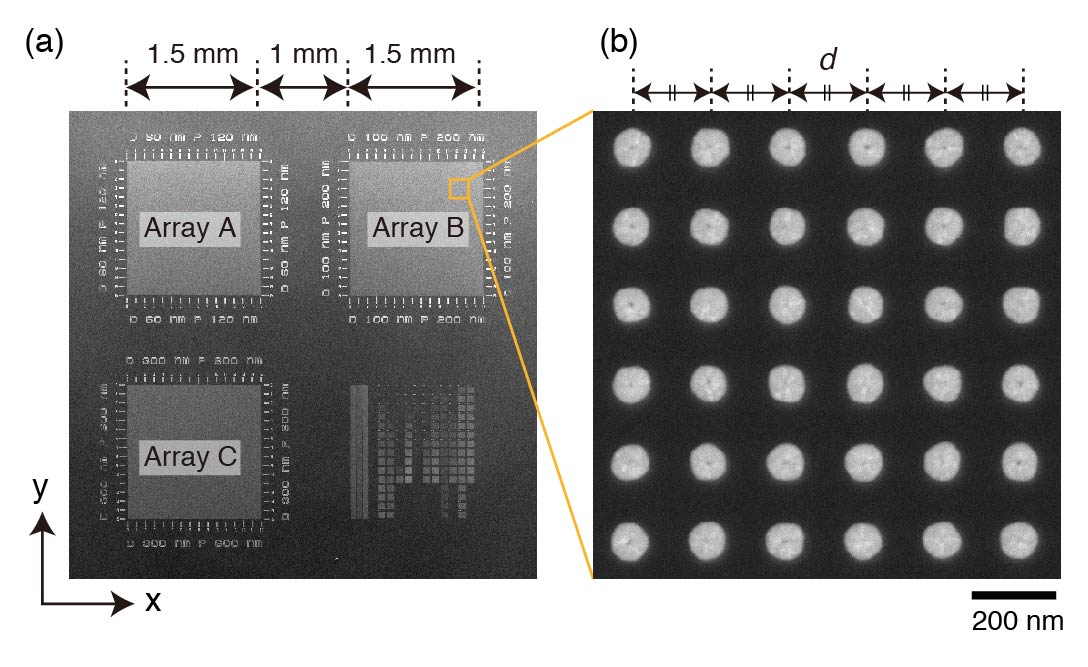
According to the procedures of DR method [1], specimens for image sharpness determination should (1) consist of particles on background (2) show enough contrast between the particles and the background (3) have no contrast anomalies such as charging effect or strong edge effect. Our candidate RM was designed to meet above three requirements and fabricated by semiconductor process [3]. As illustrated in Figure 1, this candidate RM consists of arrays of W cylinder embedded in Si substrate, which appears as dot-array in SEM images. The topographic contrast caused by the surface roughness of the candidate RM was well suppressed by making the surface flat enough via chemical mechanical polishing. The steep contrast between W dots and Si background is desirable for the image sharpness observation by DR method. In addition, with dot-pitch d, given as specified value, one can perform magnification calibration. The candidate RM has three dot-arrays, Array A, B and C, with different dot diameters (60, 100 and 300 nm, respectively) on a Si chip enabling us to choose the optimum dot size according to observing magnification.
The dot-pitch value for three arrays were SI-traceably determined by analyzing secondary electron (SE) image of the candidate RM. In every dot-pitch evaluation for three arrays, we measured 6600 dot intervals in 60 images over 10 samples. The magnification of SE images was calibrated by using SI-traceable length standard (MRS-6, Gellar MicroÅnalytical Laboratory). The dot-pitch measurement by SEM was validated by using deep-ultraviolet laser diffraction analysis that gives macroscopic view of the periodicity of the dot-array.
Obtained dot-pitch values are shown in Table. 1 with expanded uncertainty values (k = 2). The uncertainty values were evaluated by considering the homogeneity of the candidate RM, uncertainties from magnification calibration of SEM, from image analysis and from geometry of SEM observation. The predominant factor in the uncertainty was the uncertainty from magnification calibration and image digitization, which accounted more than 95% of the expanded uncertainty, while the homogeneity of the candidate RM had much small contribution to the uncertainty.
We have developed new RM that has dot-array structure, is useful for image sharpness evaluation by DR method. The dot-pitch values of three arrays were SI-traceably determined as a specified value, by which the magnification of SEM image can be calibrated with relative expanded uncertainty (k = 2) of ~1.3%.
Figure 1. SE image of the candidate RM, which consists of W dot-array embedded in Si substrate. (a) overview of the RM and (b) a magnified image of Array B.
Table 1. Dot-pitch and expanded uncertainty (k = 2) values of the candidate RM
Array Direction Dot-pitch (nm) Expanded uncertainty
(k = 2) (nm)Array A x 119.0 1.5 y 119.0 1.5 Array B x 199.1 2.4 y 199.1 2.4 Array C x 597.7 7.3 y 597.7 7.3 - References
[1] ISO/TS 24597:2011 Microbeam analysis — Scanning electron microscopy — Methods of evaluating image sharpness, (2011).
[2] M. Matthews and J. Shah, Microsc. Microanal. 21 (S3), (2015), p. 2239.
[3] K. Kumagai and A. Kurokawa, Microsc. Microanal. 22 (S3), (2016), p. 448.
[4] K. Kumagai and A. Kurokawa, Proc. MC 2017, (2017), p. 513.
[5] National Metrology Institute of Japan, NMIJ CRM 5207a Tungsten Dot-array https://unit.aist.go.jp/nmij/english/refmate/crm/certificate_sds/20/5207a_en.pdf
[6] A part of this study was conducted as one of the activities in Japan Research Industries and Industrial Technology Association. The authors are grateful to the staffs in super clean room facility (SCR) in AIST for their kind technical support in the fabrication of the candidate RM.