Challenges and perspectives of Transmission Kikuchi Diffraction in the SEM
- Abstract number
- 1124
- Event
- European Microscopy Congress 2020
- DOI
- 10.22443/rms.emc2020.1124
- Corresponding Email
- [email protected]
- Session
- PST.5 - Diffraction techniques and structural analysis
- Authors
- Dr. Alice Fanta (1)
- Affiliations
-
1. DTU Nanolab
- Keywords
Transmission Kikuchi Diffraction, improving spatial resolution, Kikuch and spot pattens
- Abstract text
Transmission Kikuchi diffraction (TKD) in the scanning electron microscope is maturing as an alternative technique to investigate crystal orientation of nanocrystalline materials. In the past few years, several applications of the technique were demonstrated1–3 and some hardware developments4,5 to improve the speed and quality of the data acquired were proposed. Furthermore, studies focusing on for example spatial resolution6, depth resolution7,8, diffraction contrast9 and comparing TKD with precession electron diffraction (SPED) in the TEM were presented10,11
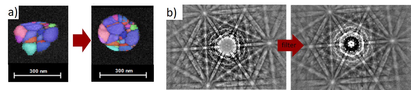
As the interest in applying TKD for nanocrystalline orientation mapping increases, it becomes important to discuss some of its challenges. In this presentation, we will first focus on describing challenges and solutions to reduce sample charge, drift and contamination during high-resolution mapping and continue by showing an approach to improve the lateral spatial resolution of TKD. Finally, we will present the perspectives of blocking the saturated signal of the transmitted beam to obtain diffraction patterns containing high contrast Kikuchi band and spot patterns. Figure 1a) shows the charge reduction achieved by investigating nanoparticles in low vacuum condition and figure 1 b) Kikuchi and spot pattern obtain from a 20nm Au film after adjusting the dynamic range of the TKD detector with a in-house developed filter.
Figure 1a) Charge control using low vacuum and b) filtering the saturated signal to obtain Kikuchi and spot patterns simultaneously.
- References
1. Alekseeva, S. et al. Nat. Commun. 8, 1084 (2017).
2. Daly, L. et al. Geochim. Cosmochim. Acta 216, 42–60 (2017).
3. Lederer, M. et al. Appl. Phys. Lett. 115, 222902 (2019).
4. Fundenberger, J. J. et al. Ultramicroscopy 161, 17–22 (2016).
5. Fanta, A. B. S. et al. Ultramicroscopy 206, 112812 (2019).
6. Niessen, F., Burrows, A. & Fanta, A. B. da S. Ultramicroscopy 186, 158–170 (2018).
7. Brodu, E. & Bouzy, E. Microsc. Microanal. 23, 1096–1106 (2017).
8. Liu, J., Lozano-Perez, S., Wilkinson, A. J. & Grovenor, C. R. M. Ultramicroscopy 205, 5–12 (2019).
9. Brodu, E., Bouzy, E. & Fundenberger, J.-J. Ultramicroscopy 181, 123–133 (2017).
10. Robert, D. et al. ACS Nano 7, 10887–94 (2013).
11. Mariano, R. G., Yau, A., McKeown, J. T., Kumar, M. & Kanan, M. W. ACS Omega acsomega.9b03505 (2020) doi:10.1021/acsomega.9b03505.