Correlating spontaneous and stimulated electron-photon interactions in nano-scale plasmonic near fields
- Abstract number
- 1024
- Event
- European Microscopy Congress 2020
- DOI
- 10.22443/rms.emc2020.1024
- Corresponding Email
- [email protected]
- Session
- PST.2 - Microscopy for the study of quantum effects and nano-optics
- Authors
- Matthias Liebtrau (1), Murat Sivis (5), Armin Feist (5), Nicolas Pazos-Pérez (4), Ramon Alvarez-Puebla (3, 4), Javier García de Abajo (2), Albert Polman (1), Claus Ropers (5)
- Affiliations
-
1. AMOLF
2. ICFO
3. ICREA
4. Universitat Rovira i Virgili
5. University of Göttingen
- Keywords
CL, EELS, near field, PINEM
- Abstract text
We correlate spontaneous and stimulated interactions of 20-200 keV electrons in scanning (transmission) electron microscopy (S(T)EM) with tightly confined plasmonic near fields at the tip apexes of a single chemically-synthesized Au nanostar, yielding new insights into the relation between electron energy loss spectroscopy (EELS), cathodoluminescence (CL) spectroscopy and photon-induced near-field electron microscopy (PINEM).
Similar to an ultrashort optical pulse, the time-varying evanescent electric field associated with a fast electron can couple with a broad spectral range of electromagnetic modes in a material [1,2]. The spontaneous energy transfer during this interaction process causes the electron to experience a characteristic energy loss that can be measured experimentally (EELS). Additionally, light emission upon radiative relaxation of the excited material can be detected in the far-field (CL). The spatial and spectral characteristics of the material resonances are directly reflected in the measured energy loss and photon emission probabilities, rendering the electron a highly versatile near field probe [3]. In PINEM, fast electrons exchange energy with an optical near field generated by an intense laser. The resulting energy-gain and -loss transitions by stimulated absorption and emission of photons cause the energy spectrum of the transmitted electrons to split into sidebands separated from the incident energy by integer multiples of the photon energy [4,5]. The population of these sidebands varies with the intensity of the laser-induced optical field, enabling spatially-resolved near field measurements similar as in EELS and CL.
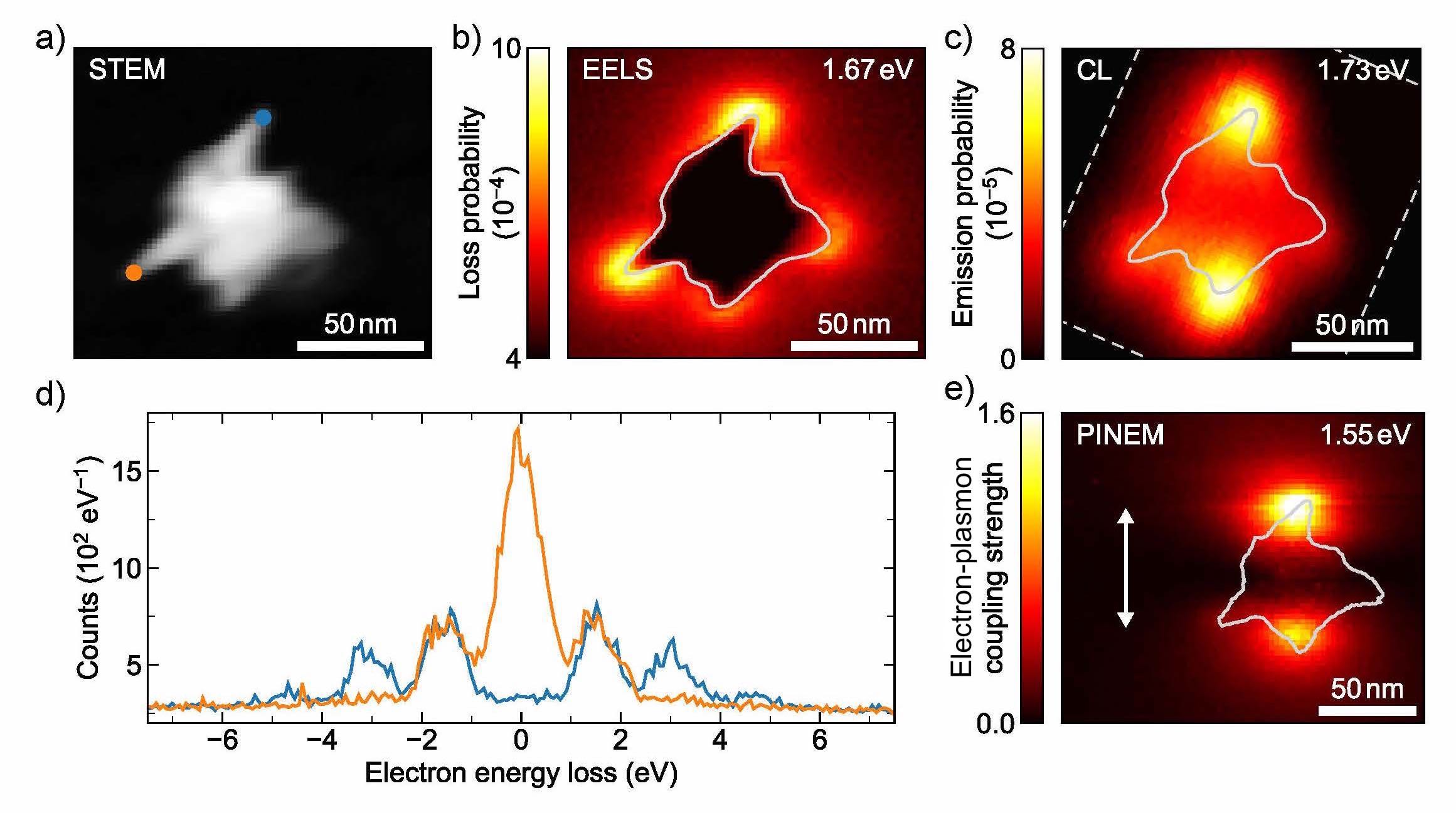
Figure 1. a) STEM bright field image of the plasmonic Au nanostar studied in this work. Near field distributions obtained from energy-filtered b) 200 keV EELS and c) 20 keV CL measurements with a bandwidth of 200 meV around the central energies indicated on the top right of each panel. d) 200 keV PINEM spectra acquired at two different tips of the nanostar indicated by the blue and orange dots in a) using 3ps, 800 nm pulsed laser excitation. e) Measured PINEM electron-plasmon coupling strength, with the white arrow indicating the laser polarization.
In this work, we demonstrate spatially-resolved EELS, CL and PINEM measurements of the plasmonic near field of a single chemically-synthesized Au nanostar with sharp elongated protrusions, featuring tip-radii of curvature smaller than 3 nm [6]. A STEM bright field image taken during EELS acquisition is shown in Fig. 1a). In good agreement with complementary boundary element method (BEM) calculations, the EELS, CL and PINEM data show that the tips of the nanostar sustain distinct dipolar surface plasmon resonances, giving rise to highly localized optical fields at the tip apexes. Energy-filtered EELS and CL distributions are shown in Fig. 1 b) and c), respectively, for a bandwidth of 200 meV around the specific energies indicated on the top right of each panel. In Fig. 1 d) we present characteristic PINEM spectra acquired at two different tips as indicated by the blue and orange dots in Fig. 1a). The spectra correspond to regions of strong (blue) and weak (orange) electron-plasmon interactions, respectively. Data reveal that for strong interaction the zero-loss peak is fully depleted and the electron energy distribution is dressed with first-, second- and third-order photon emission and absorption peaks. A PINEM near field map, derived from the energy spectra of the transmitted electrons, is shown in Fig. 1e), representing the spatial variations in the electron-plasmon coupling.
We highlight similarities and differences between the measured EELS, CL and PINEM distributions and discuss the role of the different excitation mechanisms driven either by the electron itself (EELS and CL) or the external laser (PINEM). We show that thanks to the broadband optical excitation in EELS and CL, the contribution of modes with spatial and spectral overlap can be disentangled, while in PINEM, individual tip resonances can be selectively addressed by adjusting the photon energy and polarization state of the driving field. From all three techniques, we consistently find the near field of the tip apexes to extent by only a few nanometers into the surrounding vacuum. The spontaneous and stimulated interactions of free electrons with a localized optical field reflect only a single spatial frequency component, determined by the electron velocity. As a result, electrons in the 1-30 keV SEM energy range are primarily sensitive to the most tightly confined plasmonic near fields, while electrons in the 100-300 keV STEM energy range interact more strongly with other extended modes, as we will discuss in detail.
- References
[1] F. J. García de Abajo, Rev. Mod. Phys. 82 (2010) p. 209.
[2] A. Polman, M. Kociak, and F. J. García de Abajo, Nature Mater. 18 (2019) p. 1558.
[3] A. Losquin, and M. Kociak, ACS Photonics 2 (2016) p. 1619.
[4] B. Barwick, D. J. Flannigan, and A. H. Zewail: Nature 462 (2009) p. 902.
[5] A. Feist et al, Nature 521 (2015) p. 200.
[6] N. Pazos-Pérez, L. Guerrini, and R. A. Alvarez-Puebla, ACS Omega 3 (2016) p. 17173.