Degeneration of Li-Ion batteries studied with a Field Emission Scanning Electron Microscope equipped with a Secondary Ion Mass Spectrometer
- Abstract number
- 234
- Event
- Virtual Early Career European Microscopy Congress 2020
- DOI
- 10.22443/rms.emc2020.234
- Corresponding Email
- [email protected]
- Session
- PST.3 - New Instrumentation
- Authors
- Gudrun Wilhelm (1), Dr. Ute Golla-Schindler (1), Dr. Graham Cooke (2), Dr. Timo Bernthaler (1), Dr. Gerhard Schneider (1)
- Affiliations
-
1. Aalen University - Materials Research Institute
2. Hiden Analytical GmbH
- Keywords
FIB-SIMS, Lithium-ion batteries, SEM
- Abstract text
Combined Scanning Electron Microscopy (SEM) methods obtained new insights in degradation effects occurring in the separator foil of LiB cells. The combination of SEM and mass spectrometry enables to analyze the ageing effects. The cycled separator foil contains anode residual material on top. The porous structure of the cycled separator foil contains precipitations, which restrict the pathways of the lithium ions and can cause a reduced capacity.
Commercial 18650 Graphite NMC cells represent a major part of the battery market. Due to their use in different applications the ageing processes are complex. For a long lifetime, stability in each case of use is required. Ageing mechanisms are not completely understood and a better comprehension of the ageing effects can lead to an increased battery lifetime.
The following investigations were performed on a Zeiss Crossbeam 540 with Gemini II column and a Schottky field emission electron gun. The SEM is equipped with an Everhart-Thornley detector (ETD), a backscattered electron (BSE) 4 quadrant semiconductor detector and two Inlens detectors. Cross sections can be obtained by using the Zeiss Capella focused ion beam (FIB) with a gallium liquid metal ion source. In combination with a multi gas injection system (GIS) platinum can be deposited to protect the surface. Additionally, a new Hiden Secondary Ion Mass Spectrometry (SIMS) detector consisting of a quadrupole detector can be used for chemical analysis. This combination enables the detection of light elements simultaneously with conventional SEM imaging and analyzing methods. Conventional analytical methods (like EDS) combined with SEM are restricted to the detection of elements with an atomic number above B. For the study of Li-Ion batteries the detection of lithium is important and can be realized by applying mass spectrometry, where Li can be detected regardless of the bonding situation. The SIMS detector works in combination with the FIB, which induces different effects by interacting with the bulk material. The appearance of secondary electrons and ions (partially charged) is one of the mentioned effects. The secondary electrons contain a topographical and material contrast information. The charged ions can be detected and measured in the mass spectrometer with high lateral resolution. This enables to study the chemical composition of the sample. The combination of the methods allows a new approach to analyze the samples.
The investigations were performed on two commercial 18650 cells. The cathode material is LiNi1/3Mn1/3Co1/3O2 (NMC), the anode consists of graphite. The separator foil is placed between the cathode and anode site to insulate both electrically. The cell works with a nominal capacity of 1500mAh and a cycle window of 2.7V < V < 4.2V. For our studies we use two cells, a reference cell and a cycled cell. The two cells were opened and separated into the different components (cathode, anode and separator). For the first investigation the separator foil with the side pointing to the anode was chosen. The separator foil was prepared with conductive carbon paste on SEM aluminium holder. A conductive surface coating had been left out to obtain good chemical analysis. Due to the low conductivity of the separator foil, we use low voltages and low beam currents. The images in Figure 1 and 3 were obtained at 1kV with the Inlens detector. Figure 2(c,d) and 4(a,b) were collected at 5kV using the BSE and the Inlens detector.
Between the anode material and the separator, the solid electrolyte interface (SEI) is formed by the first charging process and undergoes dynamic changes with further cycling [1]. On the cathode side, the ageing leads to the formation of the cathode electrolyte interface (CEI) [2], [3]. During the charging/discharging process, (lithium) ions cross the separator membrane which enables electrochemical reactions. The electrolyte covers anode, cathode and separator and assures ion conductivity. It is known that the ageing of the electrolyte leads to decomposition and to formation of parts of the SEI [4], [5].
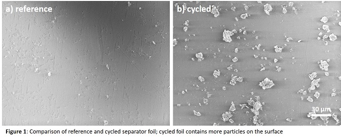
The investigation starts comparing the separator foil of reference and cycled cell at low magnification [Figure 1]. On top of the cycled separator foil, many particles and parts of the anode can be seen. The reference separator foil stays nearly free of particles. Both, size and quantity of the particles are higher on the cycled cell.
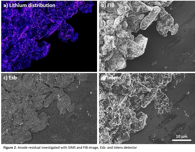
Figure 2 shows a large anode particle on top of the cycled separator foil. The images are collected with Sesi (FIB induced secondary electrons) [b], BSE [c] and Inlens [d] detector. The element distribution can be analyzed with SIMS. Exemplarily lithium distribution is shown in Figure 2a.
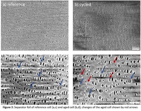
With a higher magnification the porous network structure of the separator foil is visible [Figure 3]. The regular oval shaped holes, which show an internal column structure are ordered in a sort of wavelike rows. The network structure contains local changes and seems to continue similar to the depth [blue arrows]. In Figure 3(b,d) the surface morphology of the cycled cell can be seen. Some holes are reduced in size, some are closed completely [red arrows]. The progression to the depth isn’t clearly visible but seems to show the same effect [blue arrows].
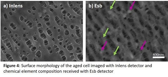
Comparing the topography (Inlens) and the material contrast (BSE) of the cycled separator foil in high magnification, the material in the closed holes could be investigated [Figure 4]. Sites with higher atomic number in the BSE image appear brighter. Two types of changes of the network structure were found. Some holes are covered with a film which appears bright [green arrows] and some holes contain agglomerations which also appear bright [pink arrows].
Comparing the reference and the cycled separator foil, three ageing effects could be detected. The quantity and size of anode material on the separator increases for the treated cell in comparison to the reference cell. Applying mass spectrometry enables the residuals of anode material on top of the separator foil to be analyzed. Exemplarily detection of lithium is presented. The porosity of the separator foil is reduced through precipitations and covering films. This effect can decelerate the lithium ion movement through the separator foil and reduce the capacity of the cell.
- References
[1] E. Peled et S. Menkin; Electrochemical Society 164 (2017) p. 1703-1719
[2] K. Edström, T. Gustafsson et J.O. Thomas; Electrochimica Acta 50 (2004) p. 397–403. DOI: 10.1016/j.electacta.2004.03.049.
[3] X. Mönnighoff et al.; Journal of Power Sources 352 (2017) p. 56–63. DOI: 10.1016/j.jpowsour.2017.03.114.
[4] E. Peled et al.; Journal of Power Sources 97-98 (2001) p.52-57
[5] P. Verma, P. Maire, P. Novák; Electrochimica Acta 55 (2010) p. 6332–6341. DOI: 10.1016/j.electacta.2010.05.072.