EBSD of granitic quartz exposed to electric field
- Abstract number
- 307
- Event
- European Microscopy Congress 2020
- DOI
- 10.22443/rms.emc2020.307
- Corresponding Email
- [email protected]
- Session
- PSA.6 - Geological Materials & Bio-mineral systems
- Authors
- Ruben Bjørge (2), Bjørn Eske Sørensen (1), Håkon W. Ånes (1), Per Erik Vullum (2), Alex Kane (2)
- Affiliations
-
1. Norwegian University of Science and Technology
2. SINTEF
- Keywords
piezoelectricity, rock, SEM
- Abstract text
Quartz is a piezoelectric mineral found in many types of rocks. When a stress is applied to a piezoelectric crystal the electric polarization is changed, while an applied electric field can induce a strain in such a crystal. Piezoelectricity is an anisotropic property: the magnitude of the induced strain depends on the crystal orientation with respect to the direction of the external electric field. The motivation for this study was to investigate if an electric field applied to a rock with high quartz-content could modify the quartz microstructure.
In this study, granite was exposed to an alternating electric field. We investigate the effect on the microstructure using scanning electron microscopy electron backscattered diffraction (SEM EBSD) of exposed and unexposed samples. Two discs of diameter 20 mm were cut out from a core of Kuru Grey granite (composed of approx. 35% quartz, the rest mainly feldspars and a small amount of mica). The discs were polished down to a thickness of approximately 1.5 mm. One sample was exposed to an alternating electric field (1 kHz) for 10 minutes by placing it between two electrodes connected to a voltage supply. The voltage was chosen such that the field was 2 kV/mm. Unexposed and exposed discs were then polished for EBSD and coated with a thin layer of carbon. SEM was performed on a Hitachi SU-6600 SEM with a Nordif EBSD system. The step size used was 2 µm. EBSD patterns were indexed using TSL OIM software. The orientation data was then analysed using MTEX, which also allows calculation of piezoelectric properties based on crystal orientation [1].
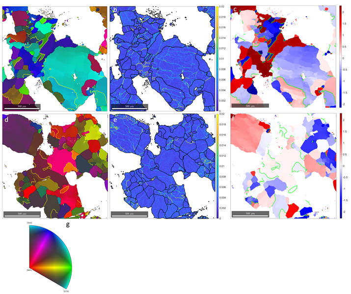
Inverse pole figure maps are shown in Figure 1(a,d) of the unexposed and exposed granite sample. Only the quartz is shown. In both samples, the quartz consists of large grains with internal deformation, and smaller strain-free recrystallized grains. This can be seen in the kernel average misorientation (KAM) maps in Fig. 1(b,e). Dauphiné twins are also found in both samples. There is no discernible difference in the level of local misorientation in the two samples.
In Fig. 1(c,f) we plot the piezoelectricity normal to the sample (along the direction of the applied electric field), as well as the Dauphiné twin boundaries. In the unexposed sample, twins appear in grains with both high and low piezoelectricity. In the exposed sample however, the twinning is mainly in grains with orientations that correspond with low piezoelectric coupling to the electric field. This might suggest that twin boundaries in grains with high piezoelectric response migrate and disappear due to the stresses induced by the applied electric field. This would be similar to removing twins from quartz crystals by applying stress [2].
Figure 1. (a-c) Inverse pole figure, KAM, and piezoelectricity map of unexposed granite. (d-f) Inverse pole figure, KAM, and piezoelectricity map of granite exposed to an alternating electric field. Dauphiné twin boundaries are indicated by yellow lines in (a, d), and green lines in (c, f). (g) Stereographic triangle for quartz to interpret colours in (a) and (d).
- References
[1] D Mainprice et al, Geological Society, London, Special Publications 409(1) (2015), p. 223.
[2] LA Thomas and WA Wooster, Proc. R. Soc. Lond. A 208 (1951), p. 43.
[3] The authors gratefully acknowledge funding from the Research Council of Norway under grant number 280755.