Electron-driven photon sources for spectral interferometry with electron microscopes
- Abstract number
- 635
- Event
- European Microscopy Congress 2020
- DOI
- 10.22443/rms.emc2020.635
- Corresponding Email
- [email protected]
- Session
- PST.2 - Microscopy for the study of quantum effects and nano-optics
- Authors
- Prof. Dr. Nahid Talebi (2), Dr. Mario Hentschel (1), Prof. Dr. Harald Giessen (1)
- Affiliations
-
1. 4th Physics Institute and Research Center SCoPE, University of Stuttgart,
2. Institute of Experimental and Applied Physics, Christian Albrechts Universty
- Keywords
Spectral Interferometry, Cathodoluminescence, Plasmons, Electron-driven photon source, Ultrafast dynamics
- Abstract text
Summary- Here, we discuss our recent achievements for the realization of electron-driven photon sources for spectral interferometry with electron microscopes. Particularly, photon-sieve-based sources with the ability to structure the light, i.e., generating light with certain radial and angular distributions, are investigated. These sources will have applications in coherent control of nanophotonics systems with electron microscopes.
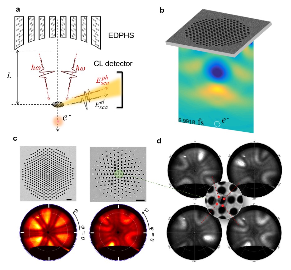
Introduction-Electron energy-loss and cathodoluminescence (CL) spectroscopy techniques are routinely employed for characterizing plasmonic materials and nanooptical excitations with high spatial resolution. By the advent of ultrafast electron microscopy, investigating the dynamics of nanooptical excitations such as plasmon polaritons have become also possible [1-3]. In these methods, a combination of electron pulses and laser pulses are used within a pump-probe spectroscopy apparatus, where the lasers are used to pump the sample to certain photonic states and electrons are used to probe the photon-induced excitations. Recently, an alternative method has been proposed, where the laser illumination is substituted by an electron-driven photon source (EDPHS) to be inserted inside an electron microscope [4] (Fig. 1a). This method has the advantage of preventing the requirements for manipulating electron microscope chambers for being adapted to the photoemission sources and the laser excitation. Also, by employing coherent mechanisms of radiation like plasmon-induced radiation, time resolution for ultrafast microscopy can be improved, due to the amelioration of the synchronization between electron and photon pulses. Moreover, thanks to the improved mutual coherence between photonic excitations of the sample and the EDPHS, interferences between these two photon excitation channels can be used for spectral interferometry with electron microscopes. Here, we outline our efforts towards the realization of a photon-sieve -based EDPHS, with tailored mechanisms of radiation.
Methods/Materials- For the realization of an EDPHS suitable for spectral interferometry, photon sieves and metamaterials design principles are employed. Our structures are comprised of an array of nanoholes incorporated into a thin gold film deposited on a Si3N4 TEM membrane (Fig. 1b), using focused ion beam. The positioning of the holes are designed in such a way, to be able to either focus the radiation onto the sample, or to structure the light to possess certain shape or directionality (Fig. 1b) [5]. We backup our experimental observations with finite-difference time-domain calculations including a relativistic charge distribution as the source. In order to characterize our designed photon sources, we have used angle-resolved CL imaging and microscopy using a Delmic SPARC detector installed in a Zeiss SIGMA field-emission microscope.
Results and Discussions- SEM images of the fabricated structures are shown in Fig. 1c. The structures hold either 4-fold or 6-fold symmetries. The radius of the holes for the 6-fold symmetric structure is uniform, and is equal to 150 nm. For the structure with 4-fold symmetry, the radius of the holes changes from 300 nm to 50nm as shown in the figuer. Angle-resolved CL maps of the structures are shown as well in Fig. 1c, demonstrating the same 4-fold and 6-fold symmetries for the structures respectively. Moreover, the angular distribution of the emitted light can be intuitively calculated based on a simple algorithm, modeling the holes as sources with induced magnetic moments (not shown here). Also, the directionality of the emission can be nicely tailored via electron impact position (Fig. 1d).
Conclusion- We have demonstrated mechanisms for designing proper electron-driven photon sources for spectral interferometry with electron microscopes, to realize structured light sources with tailored angular distributions.
Acknowledgement:
NT acknowledges support from the European Research Council (Starting Grant NanoBeam). MH and HG acknowledge financial support from the European Research Council (ERC Advanced Grant ComplexPlas), Bundesministerium für Bildung und Forschung, Deutsche Forschungsgemeinschaft (SPP1839), and Baden-Württemberg Stiftung.
Fig. 1. An EDPHS is an engineered structure with the ability to tailor the electron-induced radiation properties, such as its polarization, focal point, and direction. (a) an EDPHS can be inserted inside an electron microscope for combined electron-photon spectroscopy and spectral interferometry. (b) A photon-sieve based EDPHS for focusing the radiation at the distance of approximately 5 mm below the sample. (c) Top: SEM images, and Down: Angle-resolved CL maps acquired at . Scale bar is 2 mm. (d) Angle-resolved maps for electron impact positions depicted at the inset.
- References
[1] Yurtsever, A., van der Veen, R. M. & Zewail, A. H. Subparticle Ultrafast Spectrum Imaging in 4D Electron Microscopy. Science 335, 59 (2012).
[2] Feist, A. et al. Ultrafast transmission electron microscopy using a laser-driven field emitter: Femtosecond resolution with a high coherence electron beam. Ultramicroscopy 176, 63-73 (2017).
[3] Madan, I. et al. Holographic imaging of electromagnetic fields via electron-light quantum interference. Science Advances 5, eaav8358 (2019).
[4] Talebi, N. Spectral Interferometry with Electron Microscopes. Scientific Reports 6, 33874 (2016).
[5] Talebi, N. et al. Merging transformation optics with electron-driven photon sources. Nature Communications 10, 599 (2019).