Electron energy-loss spectroscopy using MerlinEM - Medipix3
- Abstract number
- 1384
- Event
- European Microscopy Congress 2020
- DOI
- 10.22443/rms.emc2020.1384
- Corresponding Email
- [email protected]
- Session
- PST.3 - New Instrumentation
- Authors
- Marcel Tencé (1), Jean-Denis Blazit (1), Xiaoyan Li (1), Matus Krajnak (2), Eduardo Nebot del Busto (2), Richard Skogeby (2), Léo Cambou (1), Mathieu Kociak (1), Odile Stéphan (1), Alexandre Gloter (1)
- Affiliations
-
1. Laboratoire de Physique des Solides, CNRS, Université Paris-Saclay
2. Quantum Detectors Ltd, R104 RAL, Harwell Oxford, OX11 0QX
- Keywords
direct detection, EELS, oxide
- Abstract text
M. Tencé1, JD. Blazit1, X. Li1, M. Krajnak2, E. Nebot del Busto2, RJ. Skogeby2, L. Cambou1, M. Kociak1, O. Stephan1, A. Gloter1
1 Laboratoire de Physique des Solides, CNRS, Université Paris-Saclay, 91405 Orsay, France
2 Quantum Detectors Ltd, R104 RAL, Harwell Oxford, OX11 0QX, United Kingdom
Here, the hybrid pixel detector, Medipix3, has been used for a direct electron detection in the case of EELS spectroscopy. Such detectors have already been used for imaging and diffraction in TEM [1] and STEM [2]. Several advantageous features are expected in the case of EELS (high sensitivity and dynamical ranges, adaptability from 30 to 300 keV electrons and zero read-out noise).
In order to be used as an EELS detector, Quantum Detectors designed a 4x1 MerlinEM detector where 4 Medipix3 chips are aligned in 4x1 configuration, resulting in a 1024x256 detector. A dedicated flange was design to attach the detector to a GATAN ENFINA spectrometer and this EELS system is mounted on a Cs corrected STEM (Nion USTEM200) operated at 60 and 100 keV.
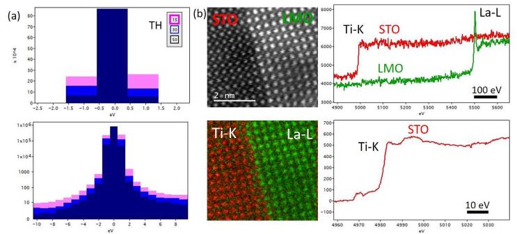
When an electron (with an Eo energy) enters in the 500 μm thick silicon layer of the Medipix3, it deposits its energy and might thus be detected by a single or several neighbouring pixels. To do so, the deposited energy must exceed the pre-set lower threshold energy value, TH, and then a count is registered in the corresponding pixel circuitry. By setting a high threshold energy, most of the electrons can only give enough energy to be detected on a single pixel (or even be undetected), resulting in a narrow point spread function (PSF). By decreasing the threshold energy, a single electron can deposit enough energy on several pixels leading to several counts and an higher detector quantum efficiency (DQE). Additionally, the detector might work in charge summing mode (CSM), where the induced current of the pixel is compared with the induced current of the surrounding pixels. Only the pixel with the highest current is thus credited of the count. This reduces the impact of charge sharing on the PSF whilst maintaining the high DQE of low threshold energies. As a result, there are several options to balance between optimizing the DQE or the PSF, that might be adapted for different EELS applications (core-loss, bang gap measurement, …) or for different electron energies Eo. Figure 1a shows two zero-loss peaks (ZLP) measured at different TH for Eo=100 keV and confirm that the PSF get better at higher TH. Nevertheless, at 100 keV for a TH = 15keV, the PSF is still of high quality with 63% of the ZLP intensity in a single pixel. Due to the high DQE, it is possible to perform spectrum imaging with atomic resolution in the range of several keV loss. Figure 1b shows the chemical map obtained on a LaMnO3 (LMO) thin film grown on SrTiO3 (STO) [3] using a 250 pA probe at 100 keV. The Ti and La atomic columns have been mapped through the Ti-K (4.9keV) and La-L (5.5keV) edges collected with 10 ms acquisition time per pixel. The maps are obtained by traditional edge subtraction on the raw data. It is also to note the advantage of “high” energy loss practiced with a rather low voltage Eo=100keV where beam damages are limited. For instance, it enables the use of larger electron doses (250pA for several minutes) for the measurement of fine structures of transition metal (TM)-K edges pre-peaks, that could be relevant to probe the local electronic structure in complement to the more established TM-L2,3 EELS edges studies (Fig 1b).
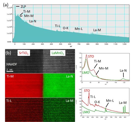
Another appealing feature of the Medipix3 is the very large dynamic range that derives from a 24-bit maximum counter depth. Every pixel can accumulate 2^24=16.8e6 counts before saturating and if the EELS spectra is spread over the non-dispersive axis, it means that a billion of counts might be obtained per energy channel. It then becomes possible to measure a non-saturated zero-loss peak, while weak signals from core-losses can be measured thanks to the intrinsic low noise level of counting detectors. Figure 2a,b show such an example for the same LaMnO3 thin film grown on SrTiO3 where the ZLP has been acquired along the core losses up to the La-M. It is thus possible to obtain a wide variety of maps (ZLP, La-N versus La-M, etc…). The difference of spatial resolution (delocalisation) between edges of low and high energy for a given element is clearly visible.
This work has received support from the National Agency for Research under the program of future investment TEMPOS CHROMATEM with the Reference No. ANR-10-EQ
Figure 1 : (a) ZLP for different threshold energies TH in linear and log scale, (b) HAADF and Ti-K, La-L EELS maps along with the EELS spectra from the STO and LMO sides. In the case of STO, EELS fine structures of the Ti-K pre-peak (4970 eV) have been measured.
Figure 2 : (a) EELS spectrum integrated from the spectrum image showing the different edges observable at the STO/LMO interface, (b) HAADF image, and the Ti and La maps obtained from the EELS spectrum image. The spectra extracted in the corresponding energy domains (20-120eV and 400-900eV) are also visible.
- References
[1] Mir et al., Characterisation of the Medipix3 detector for 60 and 80 keV electrons, Ultramicroscopy 182 (2017) 44–53.
[2] Krajnak et al., Pixelated detectors and improved efficiency for magnetic imaging in STEM differential phase contrast, Ultramicroscopy 165 (2016) 42-50.
[3] Gibert et al., Interfacial Control of Magnetic Properties at LaMnO3/LaNiO3 Interfaces, Nano Lett. 2015, 15, 7355-7361.