Evaluating brightness of pulsed electron gun using high-brightness negative electron affinity (NEA) photocathode
- Abstract number
- 549
- Event
- European Microscopy Congress 2020
- DOI
- 10.22443/rms.emc2020.549
- Corresponding Email
- [email protected]
- Session
- PST.3 - New Instrumentation
- Authors
- Hideo Morishita (2, 4), Takashi Ohshima (2), Kazuo Otsuga (2), Makoto Kuwahara (3), Yoichi Ose (1), Toshihide Agemura (1)
- Affiliations
-
1. Hitachi High-Tech Corporation
2. Hitachi, Ltd, Research & Development Group
3. Institute of Materials and Systems for Sustainability
4. Graduate School of Engineering, Nagoya University
- Keywords
negative electron affinity (NEA), photocathode, pulsed electron beam, electron microscope
- Abstract text
Recently, a novel measurement technique with a high spatiotemporal resolution that uses an electron microscope combined with a pulsed electron source has been attracting attention [1]. The photoemissive electron gun enables us to obtain an extremely high temporal resolution (typically, much less than 100 ns), which is much better than that of a conventional pulsed electron gun using a high-speed beam blanker [2]. However, the spatial resolution is restricted by the brightness of electron gun used in electron microscopes. To improve the spatial resolution, an electron source with high brightness is required.
We focus on an electron gun that uses a negative electron affinity (NEA) photocathode with p-type GaAs and Cs-O adsorbates. Under NEA condition, the bottom of the conduction band inside the GaAs is higher than the vacuum level and electrons excited from the valence band to the conduction band by light are emitted to the vacuum spontaneously. A large brightness of 1.3 × 10⁷ A/m2/sr/V was reported for an electron gun that used the NEA photocathode [3] as measured by a transmission electron microscope (TEM) [4]. This brightness is as high as that of a conventional Schottky-type emitter, which is utilized in electron microscopes with high resolution. In this research, a prototype pulsed electron gun using the NEA photocathode was constructed for a scanning electron microscope (SEM). A pulsed electron beam was obtained by irradiating the photocathode with pulsed light. The peak brightness of the beam was evaluated. We utilized two kinds of pulsed light sources with a minimum pulse width of ~100 ps and ~1 ns. In addition, we measured the waveforms of the gun with a pulse width larger than 1 ns by using a high-speed detector that consisted of an avalanche photodiode (APD) and a high-speed amplifier. In the evaluation of waveforms, pulsed electron beams with an acceleration voltage of 6 kV were irradiated on the high-speed detector, which was set in the specimen chamber of the SEM.
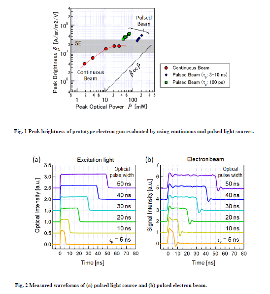
The peak optical intensity dependences of the peak brightness are shown in Fig. 1. The peak brightness of the pulsed electron beam was not saturated while that of a continuous electron beam was saturated at 2 × 10⁷ A/m2/sr/V. The maximum peak brightness was 5 × 10⁷ A/m2/sr/V, which was about 2.5 times higher than that of a conventional Schottky-type emitter. Waveforms of the pulsed light source and the pulsed electron beam are shown in Figs. 2(a) and (b), respectively. As a result, no deterioration of waveforms was observed compared with the corresponding optical waveforms. We confirmed that the prototype pulsed electron gun had an advantage in terms of generating a high-quality pulse waveform.
- References
[1] A. H. Zewail, Science 328, 187-193 (2010).
[2] S. Meuret et al., Ultramicroscopy 197, 28-38 (2019).
[3] N. Yamamoto et al., J. Appl. Phys. 102, 024904 (2007).
[4] M. Kuwahara et al., Appl. Phys. Lett. 101, 033102 (2012).