Extended 3D X-ray nanotomography of low Z and porous materials to improve statistical significance and expand electron tomography studies
- Abstract number
- 785
- Event
- European Microscopy Congress 2020
- DOI
- 10.22443/rms.emc2020.785
- Corresponding Email
- [email protected]
- Session
- DHA.2 - Advances in 3-dimensional image reconstruction
- Authors
- Silvan Englisch (1), Janis Wirth (1), Dominik Drobek (1), Dr. Benjamin Apeleo Zubiri (1), Prof. Dr. Erdmann Spiecker (1)
- Affiliations
-
1. Institute of Micro- and Nanostructure Research (IMN) & Center for Nanoanalysis and Electron Microscopy (CENEM), Friedrich‐Alexander‐Universität Erlangen‐Nürnberg, Interdisciplinary Center for Nanostructured Films (IZNF), Cauerstr. 3
- Keywords
advanced reconstruction method, correlative microscopy, segmentation, tomography, X-ray microscopy
- Abstract text
In this study, we present a method to extend the 3D volume of X-ray nanotomography (Nano-CT) studies while keeping the highest possible resolution of about 50 nm in a lab-based system ZEISS Xradia 810 Ultra. Furthermore, we demonstrate the improvement of image quality and segmentation for different low Z and porous materials. The segmentation of 3D reconstructions additionally informed by electron microscopy and electron tomography (ET) data to combine the advantages of larger volumes in X-ray microscopy and higher resolution in electron microscopy.
Microscopy, by its nature, magnifies objects and therefore focuses on rather small sample volumes. Each microscopy technique covers a specific range of sample size and spatial resolution, both being closely related. Transmission electron microscopy (TEM), in combination with ET, provides a very high resolution but is typically limited to sample areas/sizes in the micrometer range. Scanning electron microscopy (SEM), in combination with focused ion beam (FIB) tomography, enables examination of specimens at a larger scale at a slightly lower resolution but is restricted (in the case of tomography) by the destructive nature and rate of the ion milling process. At this point, lab-based X-ray microscopes such as the ZEISS Xradia 810 Ultra can extend the accessible sample volume for morphological studies to dimensions of 16 µm (HRES mode) and 64 µm (LFOV mode) at a resolution of 50 nm and 150 nm, respectively. The Xradia 810 Ultra uses monochromatic X-rays of 5.4 keV energy (Cr Ka), which for medium and high Z materials show limited penetration, often putting additional constraints on the sample size. However, for low Z and/or highly porous materials, the maximum sample size is typically not limited by absorption but by the field of view (FoV) of the X-ray optics. For these materials, we utilize Zernike phase contrast imaging to enhance material (and pore) contrast. In the first step, we present two approaches to enlarge the investigated sample size by stitching together either tilt series or tomographic 3D datasets [1]. We compare the pros and cons of the two approaches and show that the accessible sample size can be significantly increased beyond the FoV without compromising spatial resolution. In a second step, we analyze how the improved resolution affects the quality of segmentation for such larger samples. In addition, we show that the segmentation can be further improved by using complementary electron microscopy data for advanced thresholding [2].
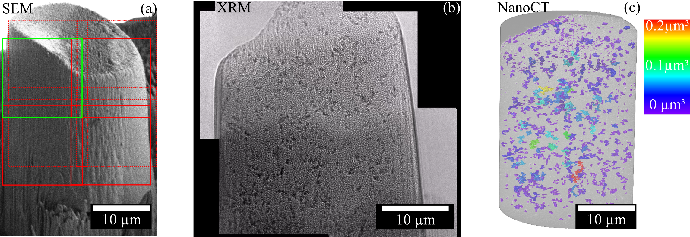
The extended tomography builds on the acquisition of multiple tilt series of overlapping sample areas (see Figure 1a). Utilizing the stage position, temperature shift tracking, and image correlation in ImageJ [3] or Python, the single images are converted to a stiched tilt series (see Figure 1b). We implement an in-house python script based on the Astra-toolbox [4] to enable the reconstruction and potentially improve it, making use of the significant number of projections and extended acquisition time. Fourier shell correlation is used to verify that the optical resolution of the HRES mode is retained in the extended tomographic reconstruction. Afterward, features of interest in the samples are segmented and analyzed according to their size, position, and spatial distribution (see Figure 1c). Depending on the sample system, the quality and reliability of segmentation are improved by employing machine learning, virtual reality analysis and electron microscopy informed thresholding.
The three investigated samples are supported catalytically active liquid metal solutions (SCALMS), MFI-type zeolite particles, and colloidal clusters made of polystyrene particles [8]. In the case of SCALMS (see Figure 1), we utilized the extended tomography to analyze the distribution of metal particles in a glass network and select features for closer analysis in TEM [5]. Regarding the MFI-type zeolites, a single macroporous zeolite particle was investigated in a correlative way by ET and Nano-CT regarding enabling improved segmentation of pore structures in Nano-CT datasets [6]. Based on this, the analysis could be extrapolated to several hundreds of zeolite particles making use of the extended tomography approach. The third investigated sample system is photonic colloidal clusters [7]. These clusters consist of primary particles with a diameter of a few hundred nanometers to achieve photonic properties in the visible or Near-IR range. By increasing the total number of these particles, the cluster sizes exceed the maximum field of view of a single ET and even Nano-CT measurements. By extending the tomography we demonstrate the capability to image these clusters up to some 100000 primary particles while preserving the maximum resolution.
In conclusion, we demonstrate the workflow and the properties of extended X-ray tomography. We utilize this method to enable a 3D investigation of samples with increased size, to improve the statistics of particle distributions in a 3D volume, to search for significant features at the range of tens to hundreds of micrometer-scale and to extrapolate the information of a single ET to extended Nano-CT datasets [8].
Figure 1. (a) Pillar sample for Nano-CT prepared from a SCLAMS sample via FIB milling. The green box corresponds to the 16 µm x 16 µm FoV of the Xradia 810 Ultra microscope, which can be imaged at 50 nm optical resolution (HRES mode). In x-, y- and z-direction tilt series are connected by an overlap (red boxes) of about 10% to extend the tomography data set volume by eight times. (b) Stiched tilt series image of X-ray projections in phase-contrast mode by ImageJ – the metal droplets appear with dark contrast embedded in the macroporous glass network. (c) Extended SIRT reconstructed Nano-CT exhibits 3D position and volume of metal particles. Greyscale threshold segmentation by machine learning in Arivis Vision4D.
- References
[1] Du, M., et al., Journal of the Optical Society of America a-Optics Image Science and Vision, 2018. 35(11): p. 1871-1879.
[2] Lenz, M., et al., Advanced Engineering Materials, 2019.
[3] Preibisch, S., et al., Bioinformatics, 2009. 25(11): p. 1463-1465.
[4] van Aarle, W., et al., Optics Express, 2016. 24(22): p. 25129-25147.
[5] Wirth, J., et al., Microscopy and Microanalysis, 2019. 25(S2): p. 422-423.
[6] Apeleo Zubiri, B., et al., Microscopy and Microanalysis, 2019. 25(S2): p. 396-397.
[7] Wang, J. W., et al., 2019. 13(8): p. 9005-9015.
[8] The authors gratefully acknowledge financial support by the German Research Foundation (DFG) within the frameworks of the research training group GRK1896 “In situ Microscopy with Electrons, X-rays and Scanning Probes”, the project SP648/8 “High-resolution X-ray microscopy for correlative tomography, high throughput screening and in situ mechanical testing of structural and functional materials” and the Collaborative Research Centre 1411 “Design of Particulate Products”. They thank P. Wasserscheid, W. Schwieger and N. Vogel and their research groups for providing the SCALMS, Zeolite and Colloidal cluster samples used in this study.