FIB-SEM and automatic segmentation for investigation of mitochondrial organization in umbrella cells of the urinary bladder urothelium
- Abstract number
- 760
- Event
- European Microscopy Congress 2020
- DOI
- 10.22443/rms.emc2020.760
- Corresponding Email
- [email protected]
- Session
- LSA.9 - Applications of volume scanning electron microscopy in life sciences
- Authors
- Rok Romih (3), Bruno Humbel (1), Manca Žerovnik Mekuč (2), Ciril Bohak (2), Matija Marolt (2), Samo Hudoklin (3)
- Affiliations
-
1. Electron Microscopy Facility, University of Lausanne
2. Faculty of Computer and Information Science, University of Ljubljana
3. Institute of Cell Biology, Faculty of Medicine, University of Ljubljana
- Keywords
dual beam, segmentation, mitochondria, urothelium, urinary bladder
- Abstract text
Introduction
Mitochondria are the main source of ATP and their ultrastructural features as well as their subcellular distribution depend on cell type and metabolism. In urothelial superficial umbrella cells, ATP is required for the synthesis and delivery of urothelial plaques by fusiform vesicles to the apical plasma membrane where they form a tight permeability barrier between urine and body fluids [1]. On the other hand, mechanical stretch of urothelium during bladder filling promotes ATP production and excretion into extracellular space [2]. ATP activates purinergic receptors on afferent nerves and interstitial cells in the lamina propria and on smooth muscle cells of detrusor. This ATP signalling pathway induces detrusor contraction and thus emptying of the bladder. Mitochondrial structural changes in urothelium were reported in aging organisms and some bladder disorders [3, 4]. Our aims were to preserve the ultrastructure of umbrella cells close to native state in the living organism and to develop a method for the automatic segmentation of mitochondria.
Materials and Methods
Mouse urothelium was high pressure frozen (Balzers HPM010), freeze-substituted (Leica AFS) and embedded in Epon. Ultrastructural features of umbrella cells were examined on ultrathin sections (Philips CM100). The volumetric data from the umbrella cells was generated by Ga-ions milling and imaging in FIB-SEM (Helios NanoLab 650). For automatic segmentation of mitochondria from FIB-SEM data, we propose a pipeline based on a volumetric convolutional neural network with mechanisms that reduce the impact of noisy and inconsistent input data. The main features of the proposed pipeline are a contrast enhancement technique, usage of segmentation masks, and zero-mean convolutions.
Results and Discussion
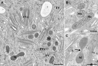
Ultrathin sections showed numerous flattened fusiform vesicles (Figure 1a) that were composed of two parallel urothelial plaques, which confirmed superior preservation of umbrella cells ultrastructure [5]. Mitochondria with smooth outer membrane were oval to elongate (Figure 1b). Sometimes narrowing was observed (Figure 1c), which point to fissions and fusions of mitochondria [3]. Our approach produced state-of-the-art results for extraction of mitochondria from FIB-SEM volumes. 3D visualization of segmented volumes presented a global distribution of numerous mitochondria within umbrella cells (Figure 2a). Some mitochondria are tubular or globular, but many of them form complex structures (Figure 2b), indicating dynamical changes possibly associated with their various functions [3, 4].
Conclusion
We believe that our approach can be used to better understand the role of mitochondria in normal and in pathological conditions in urothelium as well as other tissues.
Figure 1: Ultrastructure of the umbrella cell. A) Cytoplasm of high pressure frozen – freeze substituted umbrella cell contains mitochondria (M) and tissue specific fusiform vesicles (FV), which could also be organized into stacks (FVs). B) The shape of mitochondria on a two dimensional ultrathin section varied from being oval (Mo) to elongated (Me). C) Narrowing of the mitochondria (arrow). Bars: 500 nm – A, 250 nm – B, C.
Figure 2: Visualization of mitochondria from segmented FIB-SEM volumes. A) Mitochondria in 256×256×256 voxels. B) A part of complex mitochondria with a bifurcation and a membrane connection.
- References
[1] B Wankel et al, Mol Biol Cell 27 (2016), p. 1621.
[2] DAW Janssen, JA Schalken and J Heesakkers, Acta Physiol (Oxf) 220 (2017), p. 201.
[3] FA Kullmann et al, Neurourol Urodyn 38 (2019), p. 572.
[4] M Perse, R Injac and A Erman, PLoS One 8 (2013), p. e59638.
[5] S Hudoklin et al, PLoS One 7 (2012), p. e32935.