Graphene-molybdenum disulfide in situ mixing cells for liquid-phase transmission electron microscopy
- Abstract number
- 140
- Event
- European Microscopy Congress 2020
- DOI
- 10.22443/rms.emc2020.140
- Corresponding Email
- [email protected]
- Session
- PST.6 - In-situ and in-operando microscopy
- Authors
- Dr Daniel Kelly (1, 2), Dr Nick Clark (1, 2), Mr Mingwei Zhou (1, 2), Dr Roman Gorbachev (1, 2), Prof Sarah Haigh (1, 2)
- Affiliations
-
1. University of Manchester
2. National Graphene Institute
- Keywords
Graphene liquid cells, in situ TEM, liquid-phase TEM, two-dimensional materials
- Abstract text
In this work we present a novel design for in situ mixing of solutions within a (scanning) transmission electron microscope (S/TEM) and demonstrate its effectiveness by observing the synthesis of biomineral nanoparticles from liquid precursors in real time with sub-nanometre resolution.
Liquid-phase transmission electron microscopy (LP-TEM) is the ultimate technique for investigating dynamic chemical reactions at the nanoscale and has been facilitated by the development of environmental cells constructed from both silicon nitride (SiNx) and graphene-based membranes [1]. While silicon nitride cells have been commercialised in specialised LP-TEM holders that enable flow and mixing of liquid reagents, the resolution limit of the instrument is degraded due to the window material and liquid thickness, and mixing generally occurs outside the field of view. Flow and mixing capabilities have yet to be integrated into graphene liquid cells which conversely do allow atomic-resolution imaging of liquid-phase specimens as the electron beam is minimally scattered by the atom-thick windows and thin liquid layers [2].
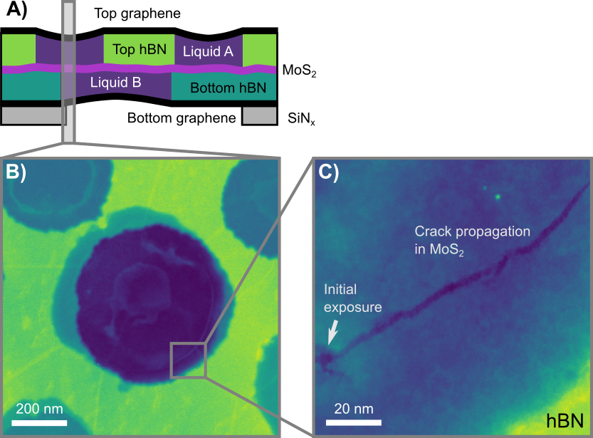
To overcome this engineering gap, we have developed a graphene mixing cell (GMC), based on previously designed engineered graphene liquid cells [3], that enables in situ mixing of two separately loaded solutions through the controlled damage of a suspended MoS2 separation layer. A schematic for this GMC is presented in figure 1A, alongside a dark-field STEM micrograph of the cell in plan-view (fig. 1B). Two etched hexagonal boron nitride (hBN) spacers are each transferred onto few-layer graphene sheets. One of these is loaded with the desired solution (liquid A) and sealed with a monolayer of MoS2, with the second graphene-hBN stack subsequently loaded (liquid B) and sealed with the graphene-hBN-MoS2 stack as illustrated in figure 1A. This results in two distinct layers of liquid cells where liquid compartments in each layer overlap to give large areas where liquids A and B are separated only by monolayer MoS2. Exposure of these areas to high electron fluence results in damage to the thin separation membrane via knock-on and radiolysis damage, and initiates mixing of the solutions.
The dark field (DF) STEM micrograph in figure 1C shows the procedure for instigating mixing in GMCs: the electron beam is positioned at a single area with high electron flux for a fixed amount of time causing a pore to form. A crack along zig-zag directions then propagates through the MoS2 layer as the lattice structure relaxes, forming a tear in the membrane 2-5 nm wide and 120 nm in length, through which liquid A and liquid B come into contact. Exploiting these nanofracture dynamics of MoS2 means that mixing can occur at a distance from the initial area exposed to high electron flux, keeping deleterious beam effects far from potential mixing sites where they are likely to influence the local reaction conditions.
We showcase this process by observing a dynamic chemical reaction whereby two aqueous solutions react to form nanoparticles via a complex biomineralisation process and confirm successful mixing by identifying the separated precursor solutions and final product using energy dispersive X-ray and electron energy loss spectroscopies (EDS, EELS). The synthesis pathways that occur as part of these biomineralisation processes have been shown to involve extremely small pre-nucleation clusters that aggregate to form crystalline particles. Due to resolution and temporal limitations, cryo-TEM has previously been used to observe freeze-frames of the reaction dynamics during early stages, while LP-TEM has been utilised to characterise the latter stages due to limited resolution and time required for pre-mixed solutions to reach the imaging area [4]. The GMCs presented here serve to overcome these limitations and provide an alternative perspective with high resolution imaging and spectroscopy as well as efficient time-scales through direct, controlled mixing within the imaging area.
These results confirm the successful mixing of two solutions within the TEM using graphene-MoS2 liquid cells, which provide advantages over traditional LP-TEM holders in that they are sufficiently thin for atomic resolution STEM imaging and also allow for direct mixing of solutions within the imaging area rather than pre-mixing in a LP-TEM holder antechamber, which has not been previously demonstrated to the best of the authors knowledge. We propose that these novel GMCs will be of great use to microscope users interested in the characteristics of dynamic chemical reactions and especially processes involving nanoscale structures, providing a complementary perspective to traditional LP-TEM holders.
Figure 1: A) Graphene mixing cell schematic showing individual components: top and bottom graphene (black), top and bottom hBN (green), MoS2 separation membrane (pink) and SiNx support grid (dark purple). B) A plan view DF-STEM micrograph where the darkest areas correspond to mixing areas (overlap of liquid wells separated by MoS2). C) High magnification STEM micrograph showing a hydrated mixing area where high electron fluence has been applied to generate a pore in the MoS2 membrane, causing a 120 nm long crack to form and leading to mixing of the liquids A and B.
- References
[1] A. S. Kashin and V. P. Ananikov, Nat. Rev. Chem. (2019) 3, 11, pp. 624–637
[2] M. Textor and N. De Jonge, Nano Lett. (2018) 18, 6, pp. 3313–3321
[3] D. J. Kelly et al., Nano Lett. (2018) 18, 2, pp. 1168–1174
[4] M. H. Nielsen, S. Aloni, and J. J. De Yoreo, Science (2014) 345, 6201, pp. 1158–1162
[5] The authors gratefully acknolwedge funding from the Engineering and Physical Sciences Research Council (EPSRC) through the Graphene NowNano Doctoral Training Centre and the European Research Council (ERC) under the European Union's Horizon 2020 research and innovation programme (ERC-2016-STG-EvoluTEM-715502)