Lorentz transmission electron microscopy of Néel-type skyrmions in multilayers of heavy metals and ferromagnets
- Abstract number
- 442
- Event
- European Microscopy Congress 2020
- DOI
- 10.22443/rms.emc2020.442
- Corresponding Email
- [email protected]
- Session
- PSA.9 - Magnetic and Spintronic Materials
- Authors
- Dr. Thibaud Denneulin (1), Dr. Jan Caron (1), Dr. András Kovács (1), Pr. Rafal Dunin-Borkowski (1)
- Affiliations
-
1. Ernst Ruska-Centre for Microscopy and Spectroscopy with Electrons and Peter Grünberg Institute, Forschungszentrum Jülich
- Keywords
Electron holography, Ferromagnetism, Fresnel, Lorentz, Skyrmions
- Abstract text
Magnetic skyrmions are foreseen as information carriers in new types of energy-efficient memories and logic devices. Multilayers of heavy metals and ferromagnets have been shown to host skyrmions at room temperature. Here, we investigate a (NiFe/Co/Pt)×5 multilayer sample using the Fresnel mode of Lorentz transmission electron microscopy (LTEM) and off-axis electron holography. We discuss how to optimize image acquisition and analysis to observe small skyrmions and compare the results with simulations.
Skyrmions are whirl-like magnetic textures that are elementary building blocks of possible future low-power storage and logic devices [1]. They can be formed at low temperature in B20-type compounds [2] and at room temperature in perpendicularly-magnetized multilayers of heavy metals and ferromagnets [3]. In such multilayers, broken inversion symmetry and a strong Dzyaloshinskii-Moriya interaction (DMI) at interfaces favors the formation of Néel-type skyrmions. In addition, the magnetic interactions can be tuned by controlling the compositions and thicknesses of the layers to obtain skyrmions of different size [4]. Magnetic skyrmions with diameters of approximately 10 nm have recently been reported [5]. Magnetic imaging techniques with high spatial resolution and sensitivity are required to investigate such small skyrmions. Although LTEM can be used to image Néel-type skyrmions by tilting the sample [6], the signal-to-noise ratio can be weak for small thicknesses of the ferromagnetic layers and as a result of diffraction contrast from the polycrystalline material. Here, we use both the Fresnel mode of LTEM and off-axis electron holography [7] to study such a multilayer sample.
The sample contains a stack of (NiFe(0.9 nm)/Co(0.2 nm)/Pt(1.5 nm))×5 deposited using DC magnetron sputtering onto a 15-nm-thick SiN membrane. Experiments were carried out at 300 kV using an FEI Titan TEM equipped with a Schottky field emission gun, a CEOS image aberration corrector, a Gatan K2 4k × 4k camera and two electron biprisms. The microscope was operated in Lorentz mode, with the first transfer lens of the aberration corrector used as the imaging lens. The objective lens was used to apply external magnetic fields to the sample.
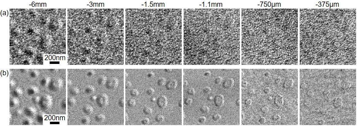
Figure 1(a) shows a defocus series of Fresnel images of individual skyrmions. The skyrmions are barely visible, even at large defocus values, as a result of the presence of local variations in diffraction contrast from the grain structure. Figure 1(b) shows the same series of images after alignment and subtraction of images recorded with the sample saturated magnetically, in order to remove non-magnetic contributions to the contrast. After subtraction, the skyrmions are more clearly visible for almost the entire range of defocus values. At larger defocus values, most of the skyrmions appear as dumbbells, with closely-spaced black and white contrast features. At lower defocus values, the edges of the skyrmions become clearer and a region of uniform intensity is visible at the centres of larger skyrmions.
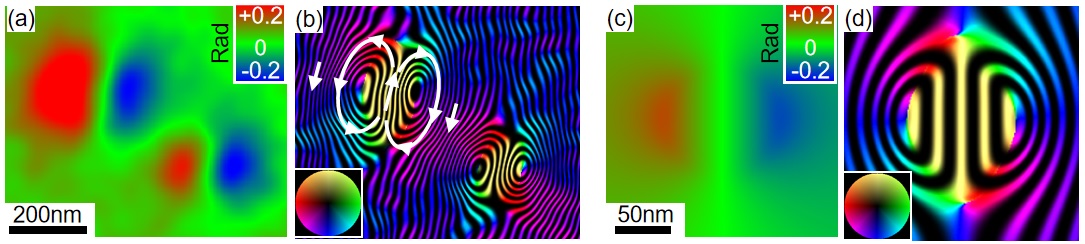
Off-axis electron holography was used to obtain more quantitative information. Part of the electron-transparent membrane was broken mechanically to create a region of vacuum for the reference wave. An electron biprism was used to overlap the reference wave with electrons that passed through the sample. Figure 2(a) shows an electron optical phase image of two skyrmions. Figure 2(b) shows the magnetic induction field obtained from the gradient of the phase image. The B field is directed upwards at the skyrmion core and downwards at the outside because of the projection of the out-of-plane component. The stray field forms two closed loops around the skyrmion core. Figure 2(c, d) shows simulations of the phase and the B field for a 100 nm Néel-type skyrmion for the experimental sample tilt and thicknesses of the ferromagnetic layers. The simulated phase variations are of the same order of magnitude as those measured experimentally.
In summary, we have used Fresnel defocus imaging to investigate a multilayer sample that hosts Néel-type skyrmions at room temperature. The magnetic contrast is weak and hidden by diffraction contrast from the crystal grain structure. Large defocus values of several mm are required to image the skyrmions, which results in blurring of the image. It is possible to improve the contrast significantly by using careful subtraction of the diffraction contrast and sub-mm values of defocus. Off-axis electron holography, in comparison with simulations, has been used to obtain quantitative phase information about the projected out-of-plane B field.
Figure 1. (a) Defocus series of Fresnel images of Néel-type skyrmions (the defocus values of -6 mm to -375 µm are indicated above the images) recorded at a sample tilt angle of 20° in the presence of an external field of 23 mT. (b) The same series after subtraction of diffraction contrast using a second series of images recorded with the sample saturated magnetically.
Figure 2. (a) Electron optical phase image of two Néel-type skyrmions recorded at a sample tilt angle of 20° in the presence of an external field of 23 mT. (b) Corresponding color-coded magnetic induction map. The contour spacing is π/100 radians. (c) Simulated phase image of a 100 nm Néel-type skyrmion for a sample tilt angle of 20°. (d) Corresponding simulated magnetic induction map.
- References
[1] A Fert et al., Nature Review Materials 2 (2017) 17031.
[2] S Mühlbauer et al., Science 323 (2009) p. 915-919.
[3] C Moreau-Luchaire et al., Nature Nanotechnology 11 (2016) p. 444-448.
[4] A Soumyanarayanan et al., Nature Materials 16 (2017) p. 898-904.
[5] L Caretta et al., Nature Nanotechnology 13 (2018) p. 1154-1160.
[6] S D Pollard et al., Nature Communications 8 (2017) 14761.
[7] A Kovács and R E Dunin-Borkowski in "Handbook of Magnetic Materials" ed. E Brück (Elsevier) p. 59-153.
[8] The authors acknowledge funding from the DARPA TEE program grant MIPR# HR0011831554.