Magnetic Field Imaging of Polycrystalline Magnets by Differential Phase Contrast STEM
- Abstract number
- 972
- Event
- Virtual Early Career European Microscopy Congress 2020
- Presentation Form
- Submitted Oral
- DOI
- 10.22443/rms.emc2020.972
- Session
- PST.1 - Phase Microscopy
- Authors
- Yoshiki O.Murakami (2), Takehito Seki (2), Akihito Kinoshita (1), Tetsuya Shoji (1), Yuichi Ikuhara (2, 3), Naoya Shibata (2, 3)
- Affiliations
-
1. Advanced Material Engineering Division, Toyota Motor Corporation
2. Institute of Engineering Innovation, The University of Tokyo
3. Nano Structures Research Laboratory, Japan Fine Ceramics Center
- Keywords
DPC-STEM, phase imaging, magnetic domain, Ned-Fe-B magnets
- Abstract text
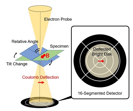
Differential phase contrast (DPC) imaging is a phase imaging technique in scanning transmission electron microscopy (STEM), which can visualize local electromagnetic field distribution inside samples in real space [1]. In DPC STEM, segmented/pixelated detectors are used to detect deflection of transmitted electron beam, which is proportional to the gradient of the phase in the transmission function (Fig.1). Combining other STEM methods such as annular dark field, energy dispersive X-ray spectroscopy and electron energy loss spectroscopy, DPC STEM can obtain electromagnetic and structural information simultaneously in the same sample area at high resolution. Thus, DPC STEM is a suitable method to investigate interaction of electromagnetic field and microstructure in materials. However, experimental DPC images contain not only electromagnetic contrast but also diffraction contrast. Diffraction contrast is so strong that we cannot distinguish between electromagnetic contrast and diffraction contrast. In single crystal, diffraction contrast can be almost eliminated by choosing a proper crystal orientation to avoid strong diffraction excitation conditions. However, in polycrystalline samples, it is almost impossible to find a specimen orientation which sufficiently eliminates diffraction contrast in all differently oriented grains.
In rare-earth magnets, the interaction between magnetic domain structures and microstructures such as grain boundary phases has been investigated for many years to improve their coercivity [2]. DPC STEM could be a promising technique to visualize magnetic fields and microstructures simultaneously if the diffraction contrast problem is overcome. It is thus desired to suppress diffraction contrast in DPC STEM images in polycrystalline samples. In this study, we show that, by averaging DPC STEM images which are acquired in slightly different tilt conditions, we can drastically suppress diffraction contrast even in polycrystalline Nd-Fe-B magnets.
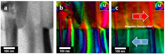
Diffraction contrast in experimental DPC images is mainly caused by a spatial intensity variation inside the bright disk. Since diffraction pattern in the bright disk strongly depends on the crystal orientation, diffraction contrast can be drastically changed by tilting the crystal orientation. We assume that the diffraction contrast is randomly varied by the sample-tilt, while the electromagnetic contrast is robust to the sample-tilt. Under this assumption, we acquired a series of DPC images in various sample-tilt conditions (Fig.1). The tilt interval was about 3 mrad and the total tilt was about 21 mrad along each tilting axis, and then totally 64 DPC images were acquired. Fig.2 (a) and (b) show an experimental DPC image of single acquisition and the averaged DPC image of the tilt-series acquisition, respectively. In the averaged DPC image, the diffraction contrast was drastically suppressed. Then we evaluated the residual diffraction contrast in the averaged image through a standard error analysis. Details of this method will be given in the presentation.
Figure. 1 Schematic image of DPC-STEM. Each segment in segmented detector acquires a signal independently.
Figure. 2 (a) Experimental ADF image of a polycrystalline Nd-Fe-B magnet. (b) Experimental DPC image. (c) The averaged DPC image of 64 DPC images acquired in the same area and various sample-tilt conditions. The magnetic easy axis of the grains is horizontal direction of images and the colour wheel with “M” represents the direction of magnetic field.
- References
[1] N. Shibata et al., Accounts of Chemical Research 50 (2017) 1502.
[2] K. Hono et al., Scripta Materialia 67 (2012) 530.