Magnetic sector SIMS system for advanced analytical capabilities on FIB instruments
- Abstract number
- 1444
- Event
- European Microscopy Congress 2020
- DOI
- 10.22443/rms.emc2020.1444
- Corresponding Email
- [email protected]
- Session
- PST.3 - New Instrumentation
- Authors
- Dr Jean-Nicolas Audinot (1), Dr Jennifer Usiobo (1), Alexander Ost (1), Charlotte Stoffels (1), Dr Tom Wirtz (1)
- Affiliations
-
1. Luxembourg Institute of Science and Technology
- Keywords
Batteries, Correlative Microscopy, Ion Beam, FIB, Photovolaic, SIMS,
- Abstract text
Techniques for nano-analysis are crucial for the ongoing investigation of nanoscale processes in disciplines from materials to life sciences. In the field of the Focused Ion Beam (FIB), the Helium Ion Microscope (HIM) has become an ideal tool for imaging and nano-patterning [1]. Imaging with helium and neon ions leads to resolutions of 0.5 nm and ~2 nm, respectively, for Secondary Electron (SE) based imaging, while structures with sub 20 nm feature sizes may be patterned using Ne. Despite these advantages, the analysis capability of the instrument is currently limited. At beam energies of 40 keV helium ions do not lead to the emission of characteristic X-rays from a sample.
Secondary Ion Mass Spectrometry (SIMS) is based on the detection of secondary ions of all elements from H to U and isotopes generated by the bombardment with an energetic primary ion beam. It’s an extremely powerful technique for analyzing surfaces, with excellent sensitivity and high dynamic range. For this purpose, we designed and developed a mass spectrometer for the HIM Orion NanoFab instrument.
This SIMS system is based on (i) specifically designed secondary ion extraction optics coupled with post-acceleration transfer optics, providing maximized extraction efficiency while keeping a finely focussed primary ion beam for highest lateral resolution, (ii) a compact floating double focusing magnetic sector mass spectrometer allowing operation in the DC mode at full transmission and (iii) a specific detection system allowing the detection of all masses in parallel. We have demonstrated that our instrument is capable of producing (i) mass spectra with mass resolution m/Δm = 400-500, (ii) very local depth profiles and (iii) elemental SIMS maps with lateral resolutions down to 12 nm [1]. Furthermore, HIM-SIMS opens the way for in-situ correlative imaging combining high resolution SE images with elemental and isotopic ratio maps from SIMS [2-5]. This approach allows SIMS maps of exactly the same zone imaged in the SE mode to be acquired easily and rapidly, followed by a fusion between the SE and SIMS data sets [5].
Here, we will review the instrument performance and present a number of examples taken from various fields of applications, ranging from materials for energy storage and production, to life sciences and geology [3-4].
- Nanoscale mapping of cathode materials of batteries was achieved by combination of SIMS and SE images. Mapping nanoscale distributions of carbon, lithium, manganese and cobalt reveals the micro-structural changes as a consequence of electrochemical reactions. Additionally, it allows identifying Li “trapping” sites within the structure that control materials properties and open the way towards designing better Li-ion cathodes.
- Correlative HIM-SIMS is also particularly well adapted for the nanoscale characterization of perovskite and CIGS solar cells. While secondary electron imaging allows the characterization of the morphology of the films, the size of the grains or the presence of defects, SIMS imaging provides the chemical information about the chemical homogeneity, the grain boundary composition and the chemical identification of nano-structures such as pinholes [3].
- In the field of life sciences, SIMS is a very interesting technique to probe labelled compounds at the cellular scale. Recently, we successfully localized the Perfluorooctanoic substances, widely used for many industrial purposes, inside the intestinal cell line (caco-2) by high-resolution SIMS. This accumulation confirms the persistence and accumulation in human and animal bodies.
- To quantify and track possible nanoscale dissolution-precipitation processes during earthquake in the presence of fluid phases, we probed on monomineralic carbonate the 1H218O water, (enriched with isotopic 18O), injected under seismic conditions. HIM-SIMS isotope mapping shows an 18O isotope enrichment only inside the fault gouge matrix.
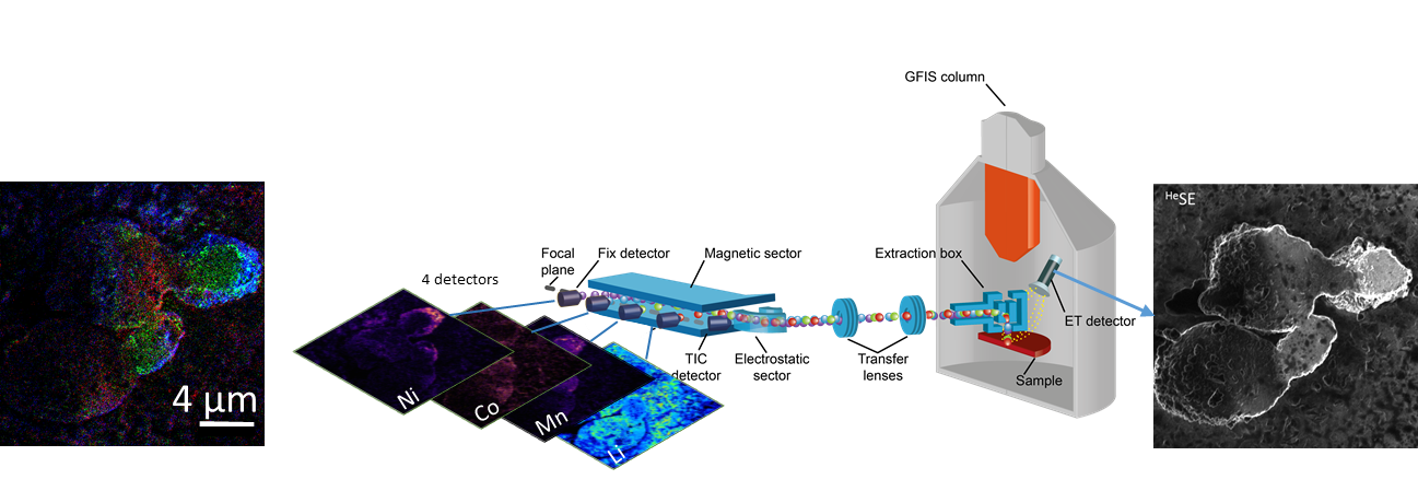
Figure 1 : Analysis of NMC battery electrode by HIM-SIMS: From right to left : Secondary Electron image (SE); schematic layout of the magnetic sector SIMS add-on system installed on the ORION NanoFab Helium Ion Microscope; SIMS images of Nickel, Cobalt, Manganese and Lithium acquired simultaneously; RGB overlay image of Cobalt (red), Manganese (Green) and Nickel (Blue). Scale bar: 4 µm.
- References
[1] D. Dowsett, T. Wirtz, Anal. Chem. 89 (2017) 8957
[2] T. Wirtz, D. Dowsett, P. Philipp, Helium Ion Microscopy, ed. by G. Hlawacek, A. Gölzhäuser, Springer, 2017
[3] T. Wirtz, O. De Castro, J.-N. Audinot, P. Philipp, Ann. Rev. Anal. Chem. 12 (2019)
[4] P. Gratia et al., J. Am. Chem. Soc. 138 (49) (2016) 15821–15824
[5] F. Vollnhals et al., Anal. Chem. 89 (2017) 10702