Nanoscale optical and vibrational spectroscopy of low-dimensional materials in electron microscope
- Abstract number
- 109
- Event
- European Microscopy Congress 2020 Invited Speakers
- DOI
- 10.22443/rms.emc2020.109
- Corresponding Email
- [email protected]
- Session
- PST.4 - Spectroscopies in Electron, X-ray and Ion Microscopy
- Authors
- Dr Ryosuke Senga (1), Dr Shigeyuki Morishita (4), Dr. Paolo Barone (5), Prof. Francesco Mauri (2, 6), Prof. Thomas Pichler (3), Dr. Kazu Suenaga (1)
- Affiliations
-
1. AIST
2. Dipartimento di Fisica, Università di Roma La Sapienza
3. Faculty of Physics, University of Vienna
4. JEOL Ltd.
5. SPIN-CNR, c/o Università G. D'Annunzio
6. Graphene Labs, Fondazione Istituto Italiano di Tecnologia
- Keywords
EELS, Vibrational spectroscopy, 1D materials, 2D materials, phonon, exciton
- Abstract text
The excitations of quasiparticles govern the physical properties of low-dimensional materials. Furthermore, assessing the irregular stacking sequences and behaviours at imperfect sections (such as boundaries and edges) is undoubtedly important to understand the performance of nanodevices. However, such local information has usually been averaged in conventional inelastic scattering techniques using x-ray, neutron, and light sources because of their diffraction limit. We demonstrate the nanoscale optical and vibrational spectroscopy of 1D or 2D materials by using a monochromatic electron source mounted in a transmission electron microscope (TEM).
All experiments were performed using a TEM (JEOL TripleC#2 at 30–60 kV) equipped with a Schottky field emission gun, a double Wien filter monochromator, and delta correctors. The energy resolution was set to a value less than 50 meV and allowed to access the quasiparticle excitations (i.e. phonon, exciton, and plasmon) of low-dimensional materials by electron energy-loss spectroscopy (EELS). The EEL spectra were collected using a GATAN GIF quantum spectrometer designed for low-voltage operations.
The spatial and momentum resolutions in electron microscopes balance each other and can be finely tuned with magnetic/electrostatic lenses. For instance, an atomically thin probe can be formed with an integration of a large momentum space. This allows us to extract local information from single defects. We have successfully measured the optical gap transitions from a defect of an individual semiconducting carbon nanotube [1,2]. Recently, atomically localized vibrational spectroscopic techniques have also been developed [3,4].
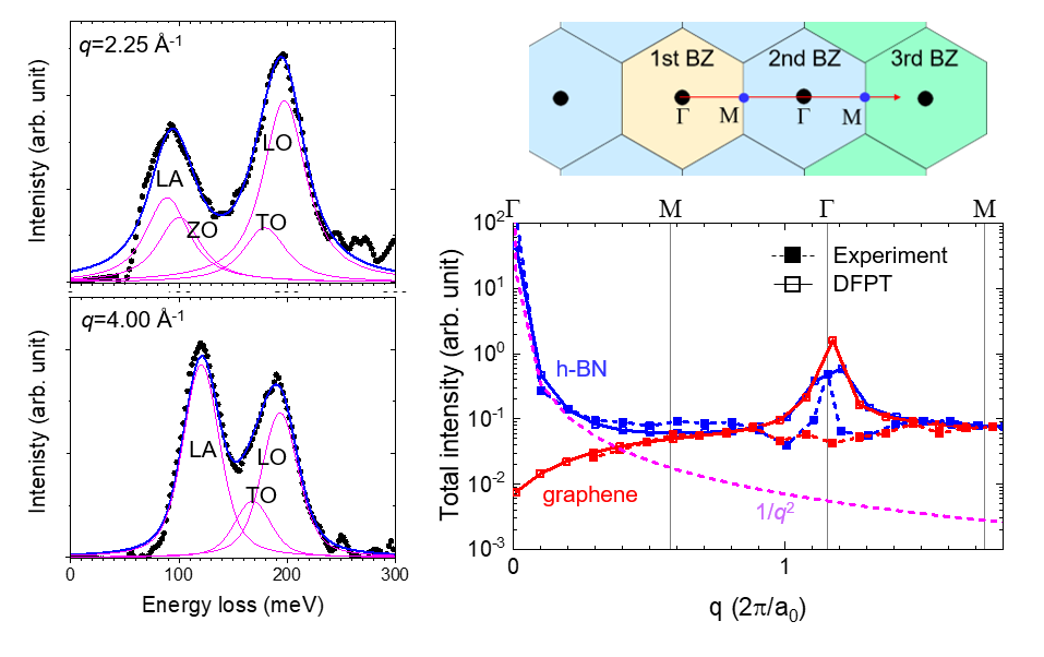
In contrast, an electron probe consisting of a parallel beam has a higher momentum resolution and provides dispersions of quasiparticle excitations. The EEL spectra are collected from each momentum transfer (q) point by a pinhole aperture in a diffraction plane. The typical EEL spectra taken from a multilayer graphene are shown in Fig. 1 (left). Using this method, we have obtained phonon dispersions of 2D materials such as graphene and h-BN [5]. Interestingly, the inelastically scattered electrons by phonon excitations provide sufficient signals at a large q even in the second and third Brillouin zone (BZ), regardless of the material polarizabilities, as shown in Fig. 1 (right). Considering charge modulations, our extended density functional perturbation theory (DFPT) fully explained the signal enhancement at a large q and accurately reproduced the measured spectra, including their intensities. Such unexpected signal enhancement at a large q helps draw a phonon dispersion curve of graphene even from a single layer. In addition, the spatial resolution of this method (a few to a few tens of nanometres, depending on its momentum resolution) is more advantageous than other dispersion measurement techniques such as inelastic x-ray spectroscopy. By maintaining the momentum resolution better than ±0.2 Å-1, each vibrational mode can be assigned at a region of a few tens of nanometres. Therefore, the propagation of each phonon mode at defects such as edges in graphene has been unambiguously visualized [5]. Such local spectroscopy with a large flexibility could help unravel the defect physics of quantum matters.
Figure 1. (left panel) The momentum-resolved EEL spectra of multilayer graphene taken at q = 2.25 and 4.00 Å-1. The EEL spectra were collected along the GMGM line which is indicated in the top-right panel. (Bottom right) The measured and simulated total intensities of phonon spectra of h-BN (blue) and graphene (red). While other quasiparticle excitations show an intensity drop following 1/q2, the phonon spectra maintain measurable intensities at large values of q.
- References
[1] R. Senga, T. Pichler and K. Suenaga Nano Letters 16 (2016), 3661.
[2] R. Senga et al., Nano Letters 18 (2018), 3920.
[3] F. Hage et al., Phys. Rev. Lett. 122 (2019), 016103
[4] K. Venkatraman et al., Nature Physics 15 (2019), 1237
[5] R. Senga et al., Nature 573 (2019), 247