Near-field Electron Ptychography using a Silicon Nitride diffuser
- Abstract number
- 66
- Event
- Virtual Early Career European Microscopy Congress 2020
- Presentation Form
- Submitted Oral
- DOI
- 10.22443/rms.emc2020.66
- Corresponding Email
- [email protected]
- Session
- PST.1 - Phase Microscopy
- Authors
- Mr Frederick Allars (2), Mr Peng-Han Lu (1), Mr Maximilian Kruth (1), Prof Rafal E. Dunin-Borkowski (1), Prof John Rodenburg (2), Dr Andrew Maiden (2)
- Affiliations
-
1. Forschungszentrum Jülich
2. University of Sheffield
- Keywords
electron microscopy, ptychography, quantitative phase imaging
- Abstract text
In this paper, we discuss a novel implementation of near field ptychography in the transmission electron microscope. The method allows a large field of view to be studied in a short time and is experimentally straightforward. We verify the precision of the method by measuring the mean inner potential of latex spheres.
Near field ptychography is an alternative phase imaging method to far field ptychography and inline holography [1]. It involves translating a sample over a grid of positions relative to structured illumination and collecting near field diffraction patterns at each point in the grid. Modulation by the diffuser results in a series of varied speckle patterns. The use of such a near field (high Fresnel number but not evanescent) method provides several advantages, including a larger field of view per diffraction pattern and reduced dynamic range compared to far field ptychography. Here, we implement near field ptychography in the transmission electron microscope (TEM) by introducing a silicon nitride diffuser in the selected area aperture plane of the microscope. We refer to this new technique as NEar field Electron pTychography, or NEET.
As NEET does not require a reference wave, unlike off-axis holography, the region of interest does not need to be close to a hole in the sample. It also does not require more than one defocus condition and inherently provides a larger field of view than either in-line or off-axis holography.
In our implementation of NEET, an FEI Titan TEM was operated in diffraction mode and the diffraction lens strength was adjusted to change the signal on the detector from a far field diffraction pattern to a defocused image of the sample. Once a diffraction condition had been obtained, a 50-µm-diameter aperture covered by a FIB-etched silicon nitride diffuser was inserted into the selected area aperture plane of the microscope. This diffuser is also defocused on the detector, resulting in interference between the two defocused fields, such that the sample transmission function is encoded by the diffuser function. A representative diffraction pattern recorded from this arrangement is shown in Figure 1b.
Once this diffraction condition is obtained, the object is moved in a grid pattern, such that successive areas of the sample that are masked by the aperture overlap. In the resulting ptychographic dataset, the ‘object’ is the image wavefront formed by the microscope objective lens, while the ‘probe’ is the transmissive silicon nitride diffuser. The data are fed into the mPIE algorithm, which recovers complex-valued images of the sample wavefront and the diffuser transmission function [2].
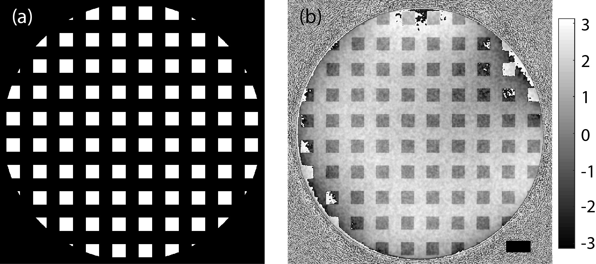
Several diffusers were designed, each providing a maximum phase shift in the incident illumination of 2.35 radians and forming speckle patterns similar to those generated by sand paper in near field X-ray ptychography [3]. A typical diffuser design is shown in Figure 2a.
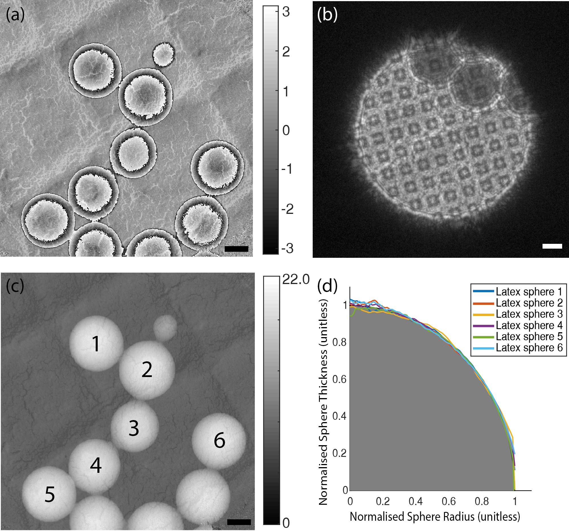
Results were obtained from a dataset consisting of a 5 by 5 raster scan of sample positions, with a step size of 100 nm. The dataset took 1 minute and 15 seconds to collect. Figure 1a shows the reconstructed phase of a test sample comprising a 500 nm diffraction grating replica populated with 261-nm-diameter latex spheres. An area of 1.5 by 1.4 µm was reconstructed with a pixel size of 1.25 nm. A Fourier ring correlation gave a ½ bit resolution of 5 nm [4].
The accuracy of the reconstructed phase in Figure 1a was assessed by using a method described by Maiden et al [5] based on the measurement of the mean inner potential of latex spheres. A value of 8.46 V was obtained with a standard deviation of 0.62 V [6]. Figure 1c shows the unwrapped phase of the latex spheres, from which the mean inner potential was measured. A plot of the radially average unwrapped phase is shown alongside the projected thickness profile of a sphere in Figure 1d. The diffuser transmission profile was also recovered during ptychographic reconstruction, and showed excellent quantitative agreement with the designed phase profile, as shown in Figure 2b.
By using the NEET method, a near-field ptychographic dataset can be collected in approximately 1 minute, whereas in our previous work the data collection time approached 1 hour [5]. Here, the resulting phase images provide a large field of view and a resolution of 5 nm. Furthermore, the phase images agree well with known values for the mean inner potential of latex spheres, providing confidence that NEET is a quantitative technique.
Figure 1: (a) Reconstructed phase of a grating replica populated with 261-nm-diameter latex spheres. (b) Representative NEET diffraction pattern. (c) Unwrapped phase of (a), which was used to measure the mean inner potential of the latex spheres. (d) Radially averaged unwrapped phase profiles of the spheres labelled in (c). The grey area corresponds to the projection of a perfect sphere, to which the phase profiles were fitted. The scale bars in (a)-(c) are 100 nm. Colour Bars in radians.
Figure 2: (a) Phase of the diffuser design. The phase difference is 2.35 radians. (b) Phase of the reconstructed square grid diffuser. The scale bar is 5 µm. The grey scale is in radians.
- References
[1] M. Stockmar et al., "Near-field ptychography: phase retrieval for inline holography using a structured illumination," Scientific reports, vol. 3, p. 1927, 2013.
[2] A. Maiden, D. Johnson, and P. Li, "Further improvements to the ptychographical iterative engine," Optica, vol. 4, no. 7, pp. 736-745, 2017.
[3] R. M. Clare et al., "Characterization of near-field ptychography," Optics express, vol. 23, no. 15, pp. 19728-19742, 2015.
[4] M. Van Heel and M. Schatz, "Fourier shell correlation threshold criteria," Journal of structural biology, vol. 151, no. 3, pp. 250-262, 2005.
[5] A. M. Maiden et al, "Quantitative electron phase imaging with high sensitivity and an unlimited field of view," Scientific reports, vol. 5, p. 14690, 2015.
[6] Y. C. Wang et al, "Measurement of polystyrene mean inner potential by transmission electron holography of latex spheres," Microscopy and Microanalysis, vol. 4, no. 2, pp. 146-157, 1998.