Quantification of volume changes during dehydration and clearing of tissue samples
- Abstract number
- 1360
- Event
- European Microscopy Congress 2020
- DOI
- 10.22443/rms.emc2020.1360
- Corresponding Email
- [email protected]
- Session
- LSA.5 - Metabolic and Large Scale Imaging
- Authors
- Barbora Radochova (3), Majda Crnak Maasarani (2), Martin Capek (3), Natasa Pollak Kristl (2), Nejc Umek (2), Jiri Janacek (3), Tatjana Stopar Pintaric (1), Erika Cvetko (2)
- Affiliations
-
1. Clinical Department of Anaesthesiology and Surgical Intensive Therapy, University Medical Center
2. Institute of Anatomy, Faculty of Medicine, University of Ljubljana
3. Institute of Physiology of the Czech Academy of Sciences
- Keywords
BABB, optical projection tomography, peripheral nerve tissue, shrinkage, stereology
- Abstract text
In this study, volume changes of the nerve tissue samples were evaluated for standard method of sample preparation/clearing with BABB (benzyl alcohol benzyl benzoate) solution. Samples were visualized using optical projection tomography (OPT) and volume changes were quantified stereologically. The volume of the samples generally shrinked by propoortion in the interval of 18 to 24%. More detailed analysis revealed proportional shrinkage of neural and non-neural tissue.
One of the pitfalls of clearing is propensity of the tissue samples to shrink or swell, depending on the method used. Standard method of methanol dehydration and BABB clearing is known to cause shrinkage of tissue samples as we demonstrated on whole mouse embryos [1]. In the present study, we used the same method for more detailed evaluation of volume changes in peripheral nerve tissue samples. The aim was to assess shrinkage of both the nerve fascicles (neural tissue) and epineurium (non-neural tissue). Samples were visualized using OPT method as we proved it to be a suitable tool to study 3D microanatomy of nerves [2].
The study was conducted on three types of peripheral nerves: n. median, n. radial and n. ulnar. Four segments of each nerve (about 3-4 mm long) were taken from autopsy specimens removed within 24 hours of death. The samples were fixed in 4% PFA, embedded into a block of agarose and scanned in a custom-made OPT scanner developed in cooperation with the Politecnico di Milano, Italy. The samples were then dehydrated through an ethanol series and cleared for 24h in BABB (1 part benzyl alcohol to 2 parts benzyl benzoate). After clearing, the scanning of the samples was repeated. For both scannings, 2x Plan-Apo Infinity-Corrected ELWD objective (Edmund Optics, USA) was used. The images were acquired using autofluorescence of the samples (excitation at 405 nm and high pass emmision filter from 550nm). Step angle was set to 0.9°, yielding 400 images per scan. Tomographic reconstructions were calculated using a filtered back-projection algorithm in NRecon software (Bruker, USA). The reconstructed data (series of optical sections) were used for estimation of the whole volume of the samples using virtual spatial grid of lines, the Fakir Method [3]. For estimation of the changes in the nerve fascicles and the epineurium, Point Grid plug-in in Ellipse software (ViDiTo, Slovakia) was used on projected cross-sections.
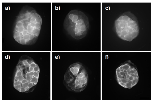
Original volumes of the individual nerve samples ranged in between 12 to 36 mm3. During dehydration and clearing procedure, the water loss caused the shrinkage of the original volume of the samples in range of 18-24%. We found no significant differences between the three types of nerves. More detailed measurement of the ratio of the neural and non-neural tissue made on projected images of cross-sections of the samples before and after clearing (Fig.1) revealed proportional shrinkage of both types of tissue. Again there were no differences between the three types of nerves. As the peripheral nerve tissue contains about 60% of water [4], some shrinkage during dehydration and clearing is to be expected and should be considered in studies dealing with peripheral nerve microanatomy.
Dehydration and clearing of peripheral nerve tissue samples with BABB caused shrinkage of about one fifth of their volume. The shrinkage was proportional for both type of tissue (neural and non-neural). There were no differences between the three types of nerves [5].
Figure1. Tomographic projections of peripheral nerves before (a-c) and after (d-f) dehydration and clearing procedure. Nerve median (a,d), nerve radial (b,e) and nerve ulnar (c, f). Bar means 1mm.
- References
[1] H Kolesová et al, Histochem Cell Biol 146 (2016), p. 141-152.
[2] A Prats-Galino et al, Clin Anat 31 (2018), p. 424-431.
[3] L Kubínová, J Janáček, J Microsc 191 (1998), p. 201-211.
[4] A Anghinah, FB Dejorge, J Aisen, Clin Chem 15 (1969), p. 1230-1233.
[5] Supported by CZ.02.1.01/0.0/0.0/16_013/0001775 Modernization and support of research activities of the national infrastructure for biological and medical imaging Czech-BioImaging funded by OP RDE, by MEYS (LM2015062 Czech-Bioimaging, LTC17023 INTER- COST) and by Slovenian Research Agency (P3-0043).