Quantitative 3D Reconstruction of Porous Polymers for Controlled Drug Release using FIB-SEM Tomography
- Abstract number
- 1448
- Event
- European Microscopy Congress 2020
- DOI
- 10.22443/rms.emc2020.1448
- Corresponding Email
- [email protected]
- Session
- LSA.12 - Application of EM in health industry
- Authors
- Cecilia Fager (2), Sandra Barman (2), Magnus Röding (3), Anna Olsson (1), Niklas Lorén (3), Johan Hjärtstam (1), David Bolin (2), Christian von Corswant (1), Holger Rootzén (2), Tobias Gebäck (2), Aila Särkkä (2), Eva Olsson (2)
- Affiliations
-
1. AstraZeneca
2. Chalmers University of Technology
3. Research Institutes of Sweden
- Keywords
3D, electron microscopy, controlled drug release, coatings, polymers, mass transport
- Abstract text
Controlled drug release is used to optimise the therapeutic efficiency of medical pellets. One way to control the drug release is to coat the medical pellet with a phase separated polymer coating. The coating consists of one water soluble polymer and one water insoluble polymer. The water soluble polymer leaches when the coating is brought in contact with body fluids and a porous structure is formed. The porous network provides transport paths for the drugs [1]. The interconnectivity of the porous network strongly influences the transport properties. The correlation between the detailed structure of the network and the transport properties illustrates the importance of quantifying the interconnectivity in 3D.
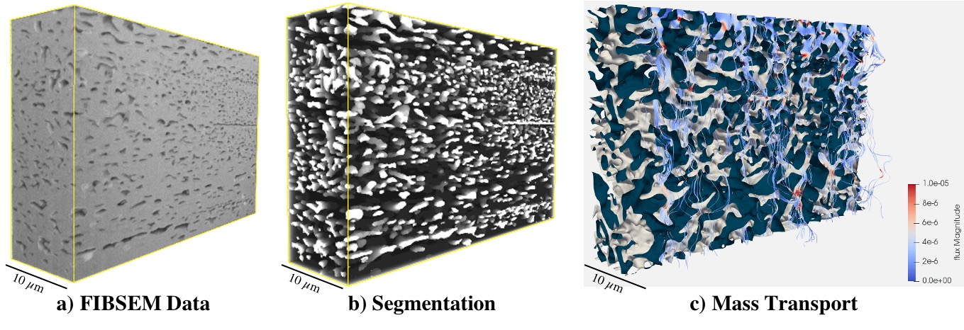
Here we present the correlation between the structure of the porous network and materials properties of phase separated polymer coatings consisting of ethyl cellulose (EC), water insoluble, and hydroxypropyl cellulose (HPC). We have acquired high spatial 3D resolution data on the porous coatings using FIB-SEM tomography, see Figure 1a). We have optimised the FIB-SEM parameters and established a generic protocol for porous and poorly conducting materials in order to overcome challenges such as redeposition, curtaining, shadowing effects, charging and sub-surface information due to the pores. In addition, a new machine-learning segmentation algorithm has been introduced to enable an automatic separation between pores and matrix, see Figure 1b). The quantification of the porous network has been carried out by determining the pore size distribution, tortuosity and interconnectivity. As a final step, diffusion simulations have been performed on the FIB-SEM data and correlated with experimentally measured permeability, see Figure 1c). [2]
Figure 1. Shows three 3D reconstructions with the volume 30 µm x 20 µm x 10 µm. a) 3D reconstruction of porous polymer film used for controlled drug release obtained using a focused ion beam with scanning electron microscope. b) segmented FIB-SEM data where the porous network is shown in white and the solid ethyl cellulose is shown in black. c) mass transport simulation performed on 3D reconstructed FIB-SEM data. The red flux lines corresponds to higher flux values while the blue flux lines to lower flux values.
References
[1] Siepmann, F.; Hoffmann, A.; Leclercq, B.; Carling, B.; Siepmann, J. How to Adjust Desired Drug Release Patterns from Ethylcellulose-Coated Dosage Forms. Journal of Controlled Release. 2007, 119, 182-189.
[2] We gratefully acknowledge financial support from the Swedish Foundation for Strategic Research (SSF), AstraZeneca for providing the materials and Chalmers Material Analysis Laboratory for technical support.