Scanning transmission imaging in the helium ion microscope with a position-sensitive detector
- Abstract number
- 995
- Event
- Virtual Early Career European Microscopy Congress 2020
- Presentation Form
- Submitted Oral
- DOI
- 10.22443/rms.emc2020.995
- Corresponding Email
- [email protected]
- Session
- PST.3 - New Instrumentation
- Authors
- Eduardo Serralta (2, 5), Nico Klingner (2), Olivier De Castro (3), Antje Biesemeie (3), Michael Mousley (3), Cecilia Bebeacua (1), Ciarán Fowley (2), Santhana Eswara (3), Serge Duarte Pinto (4), Tom Wirtz (3), Gregor Hlawacek (2)
- Affiliations
-
1. Eidgenössische Technische Hochschule Zürich
2. Helmholtz-Zentrum Dresden-Rossendorf
3. Luxembourg Institute of Science and Technology
4. Photonis Netherlands B.V
5. Technische Universität Dresden
- Keywords
Helium Ion Microscopy
Instrument development
Scanning Transmission Ion Microscopy- Abstract text
The helium ion microscope (HIM) is an instrument for high-resolution imaging, nanofabrication, composition analysis, and material modification at the nanometer scale [1]. Imaging in transmission can further improve the instrument capabilities. Mass-thickness [2], channeling [3], diffraction [4], and energy-loss are possible contrast mechanisms that can be used in transmission mode to provide further information about samples under investigation.
To study the potential of scanning transmission ion microscopy (STIM), a position- and time-sensitive detector comprising a microchannel plate and a delay line readout structure was developed. An in-vacuum linear support is used to control the detector distance to the sample. This optimization of for either maximum collection angle, or for higher angular resolution and longer time-of-flight enables, using the same system, the analysis of high-angle scattering phenomena (dark-field contrast) and the small scattering angles phenomena (such as channeling and diffraction). Post-processing the data allows the reconstruction of images for selected scattering directions and angles.
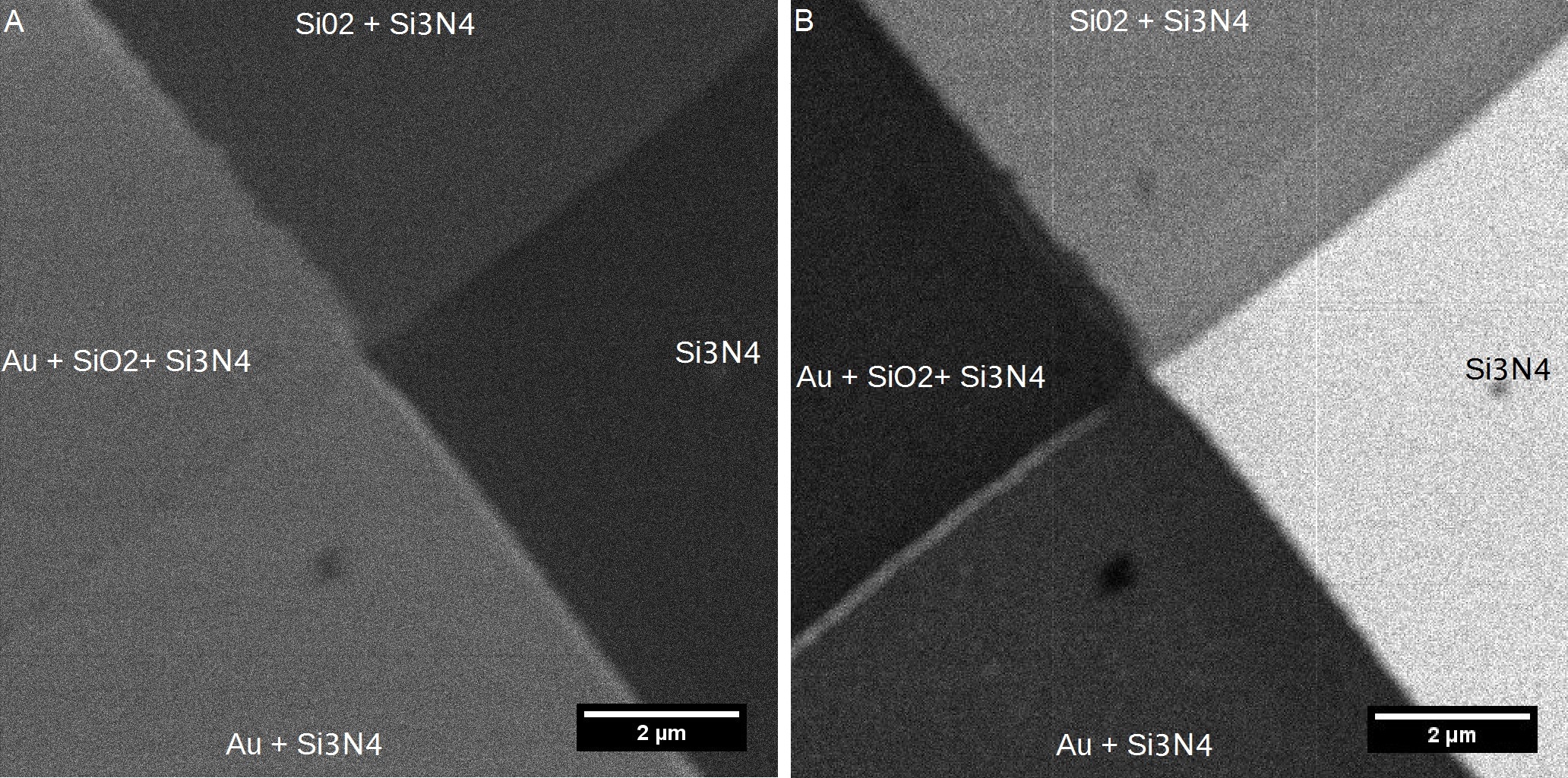
Material contrast for layered films of various materials is shown in Figure 1. Further results on other types of samples using this technique are going to be presented as well.Figure 1: Silicon nitride window with deposited layers of silicon dioxide and gold. A) Helium ion micrograph using the Everhart-Thornley detector signal. B) Bright-field STIM image of the same field of view showing contrast for each area of the sample.
- References
[1] G. Hlawacek et al., J. Vac. Sci. Technol. B 32, 20801 (2014).
[2] A. R. Hall, Microsc. Microanal. 19, 740, (2013).
[3] J. Wang et al. J. Vac. Sci. Technol. B, Nanotechnol. Microelectron. Mater. Process. Meas. Phenom. 36, 021203, (2018).
[4] J. A. Notte et al. Microsc. Microanal. 16, 599, (2010).
[5] This work has been supported by the H2020 Project npSCOPE under grant number 720964.