SPECTROSCOPIC COINCIDENCE EXPERIMENTS IN TRANSMISSION ELECTRON MICROSCOPY
- Abstract number
- 993
- Event
- European Microscopy Congress 2020
- DOI
- 10.22443/rms.emc2020.993
- Corresponding Email
- [email protected]
- Session
- PST.4 - Spectroscopies in Electron, X-ray and Ion Microscopy
- Authors
- Daen Jannis (1, 4), Knut Müller-Caspary (2), Armand Béché (1, 4), Andreas Oelsner (3), Johan Verbeeck (1, 4)
- Affiliations
-
1. EMAT, University of Antwerp
2. Ernst Ruska-Centre for Microscopy and Spectroscopy with Electrons and Peter Grünberg Institute
3. Surface Concepts GmbH
4. NANOlab Center of Excellence, University of Antwerp
- Keywords
Electron-energy loss spectroscopy
Energy dispersive X-ray spectroscopy
Electron transmission microscopy
Coindidence technique
- Abstract text
Modern transmission electron microscopes are often equipped with electron energy loss spectrometers (EELS) and energy dispersive spectrometers (EDX). Both measurement techniques share the fact that excitations of atomic states are involved. Indeed, for every X-ray emitted there was at least one electron that gave part of its energy to excite the atom in the first place, and therefore one could imagine that they convey very similar information. However, conventional EELS suffers from a large background signal and the current EDX detectors lack energy resolution. Since the two signals originate from the same process, the temporal correlation between these signals can be measured in order to select only electrons and X-rays coming from the same process hence significantly reducing the background signal. The idea of observing such temporal correlations has previously been applied by Kruit et al.[1] making use of real time event filtering and serial EELS acquisition.
In the present work, we demonstrate the capability of detecting the single electron events with a time resolution in the 100 ps range by using a novel delay line detector [2], which was placed at the end of a spectrometer, and a Super-X EDX detector setup. This setup shows the potential to improve the signal-to-background ratio of the EEL spectrum and revealing the inelastic scattered electrons coming from low abundance elements in a matrix of majority elements [3]. This is achieved by selecting only the electron events which have an X-ray detected inside the time correlation window, which is determined by the time resolution of the detection setup. This method is different from previous setups as every single event is detected, timestamped and filtering happens afterwards while still having access to the conventional EEL and EDX spectra as well.
Finally, a new setup is described where the delay-line-detector is replaced by a Timepix3 hybrid pixel detector setup[4]. This detector is able to measure individual electrons with a time resolution of 4 ns. For EDX, a digital pulse processor has been incorporated in order to improve the time-of-arrival resolution and to allow using all four detectors increasing the collection efficiency. This new setup replaces the conventional frame based signal detection into an event driven one where on top of the conventional signals, time correlations on the order of ns can be observed. We will demonstrate the latest results on this setup while speculating about its possibilities and further growth potential.
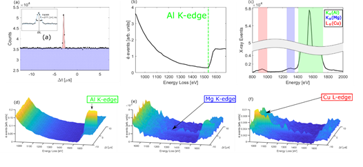
Figure 1. (a) . Histogram of the time difference ∆t between X-ray and EEL events showing a clear temporal correlation(red peak). (b) The EEL spectrum of the Al-Mg-Si-Cu alloy where the aluminium K-edge is indicated. (c) The X-ray spectrum where three X-rays originating from different atoms are marked. (d-f) The post selection of electron events, when different energy windows of X-ray energies are selected, as a function of the electron energy loss and time difference between the X-ray and electron event. The selected energy windows correspond to the characteristic X-ray energies of the different atoms (Al, Mg, Cu) present in the sample. It is clear that at a particular time difference there is an increase in signal. This increased signal corresponds to the core-level ionization event followed by X-ray emission with an energy inside the selected window.
- References
[1] P. Kruit et al. Ultramicroscopy, 13(3):205-213 (1984).
[2] A.Oelsner et al, Rev. Sci. Intrum. 72, 3968 (2001)
[3] D. Jannis et al, Appl. Phys. Lett. 114, 143101 (2019)
[4] X. Llopart et al, Nucl. Instr. And Meth. A 581 485-494 (2007)
[5] D.J., A.B. and J.V. acknowledge funding from the Flemish Research Fund FWO under project no. G093417N and G042920N, J.V. acknowledges funding from ESTEEM3, K.M. acknowledge funding from Helmholtz project no. VH-NG 1317