Structure of nanophase precipitates in La0.1Sr0.9TiO3 determined by precession electron diffraction tomography
- Abstract number
- 302
- Event
- European Microscopy Congress 2020
- DOI
- 10.22443/rms.emc2020.302
- Corresponding Email
- [email protected]
- Session
- PST.5 - Diffraction techniques and structural analysis
- Authors
- Mr. Ercin Duran (1), Dr. Feridoon Azough (1), Dr. Alexander Eggeman (1)
- Affiliations
-
1. The University of Manchester
- Keywords
precession electron diffraction, structure solution, superstructure, thermoelectrics
- Abstract text
Three-dimensional electron diffraction studies have elucidated the structure of a novel ordered phase in the La-SrTiO3 system. An overall stoichiometry of LaSr7Ti8O22 leads to a monoclinic perovskite supercell with a brownmillerite structure localised near the lanthanum substituted positions.
Nanostructured thermoelectric materials have gained attention recently due to their superior thermoelectric properties when compared to the same material produced with the classical synthesis route. The enhanced thermoelectric response is generally attributed to the nano-sized features that are formed during the synthesis and alter the electron scattering rates favourably. Understanding the crystal structure of these features is essential since they have a direct effect on the overall thermoelectric property of the material. Azough et al. recently reported on La-doped SrTiO3 with enhanced thermoelectric response due to this nanostructuring phenomenon [1]. The results indicated that the cores of the grains contain a number of nanoscale precipitates with a different crystal structure which leads to formation of a precipitation free zone around the grain boundaries. However analysis of HRTEM images was unable to discern the atomic arrangement within the precipitates. In this study, the crystal structure of the nano-crystals of La-doped SrTiO3 was investigated by using advanced diffraction techniques in TEM. The reciprocal space of a single crystal was sampled with a precession electron diffraction tilt series. The reconstructed reciprocal lattice indicated the presence of a superstructure coherent with the parent SrTiO3 perovskite.
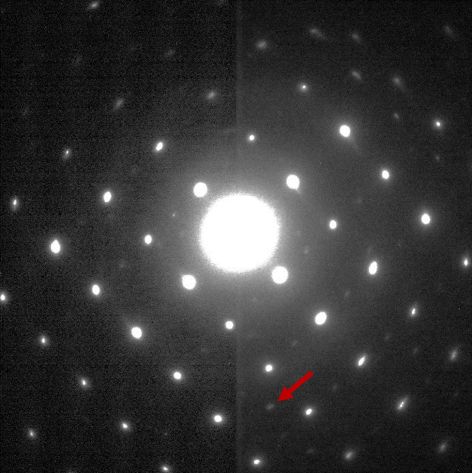
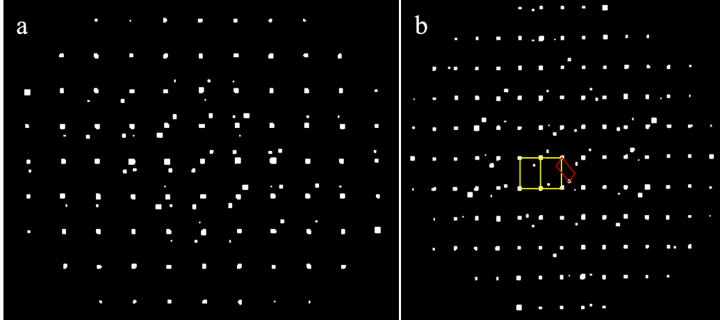
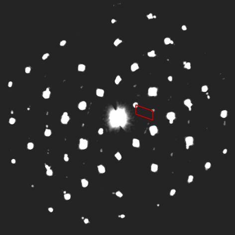
Individual diffraction patterns of a La-SrTiO3 single crystal were collected while tilting on an arbitrary axis and an example diffraction pattern is shown in Figure 1. The tomography data was analysed using the PETS software package which allowed the reciprocal lattice of the material to be reconstructed. Superlattice reflections between the reflections of the parent cubic structure indicated a coherent but much larger cell, a cubic projection of the reconstructed reciprocal lattice is demonstrated in Figure 2-a. Despite the high degree of coherency between the supercell and the perovskite, the supercell found to be a monoclinic with the parameters, 6.73 Å (~ap√3), 5.47 Å (~ap√2), 13.27 Å (~ap2√3) , and 109.2° for a, b, c, and β, respectively (where ap ~3.85 Å). Figure 2-b shows the [010*] projection of the monoclinic cell, indicating the legitimacy of the proposed cell. Precise determination of the space group is difficult with no conclusive evidence of a systematic absence in h0l reciprocal space section (Figure 3). As a consequence the lower symmetry P2 or P2/m spacegroups were used for further analysis rather than P21 or a C-centred lattice.
Figure 1. An example electron diffraction pattern of La-SrTiO3, a supercell reflection is highlighted with an arrow.
Figure 2. Reconstructed reciprocal lattice projections, (a) [001] type projection of the cubic perovskite subcell, (b) [010*] projection of the monoclinic cell with the both cells shown.
Figure 3. Reconstructed reciprocal space section, h0l plane, with the monoclinic cell shown.
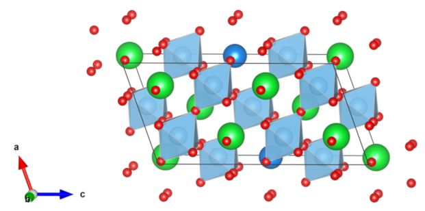
By utilising charge-flipping [2] and direct methods [3] approaches, the structure of the supercell was determined as shown in Figure 4, where green atoms are denoting Sr, red atoms O, dark blue atoms La and the blue octahedra Ti-O. Systematic structure solution attempts converged on the chemical composition of LaSr7Ti8O22, indicating an increase in La/Sr ratio as well as a deficiency in oxygen which are in good agreement with the EELS experiments of the previous study [1]. The structure accommodates the variation in stoichiometry by maintaining the general perovskite structure of the strontium titanate with a regular substitution of a lanthanum ion onto one of the strontium positions in the cell. The oxygen sub-stoichiometry manifests as a regular vacancy on an oxygen site in the first neighbour shell to the lanthanum site. This creates a brownmillerite structure [4, 5] of square pyramid coordinated titanium ions (Ti3+) close to the lanthanum site but maintains the near pristine perovskite structure through the rest of the cell.
Figure 4. The as-solved structure of La-SrTiO3 supercell
Kinematical refinement attempts produced unphysical oxygen positions; however, the oxygen deficiency was consistently attributed to two of the oxygens that are closest to La atoms. Since the superstructure is embedded in the perovskite matrix it is likely that the intensities of the supercell reflections were heavily overlapped with the perovskite subcell reflections and thus kinematical refinement was not possible.
It is conceivable that local variations in the average 10% La doping in Sr sites of SrTiO3 surpass the solubility limit of lanthanum in the strontium titanate matrix, this leads to a transition from random lanthanum positions to an ordered arrangement, these separate as widely as possible in order to minimise the distortion of the remainder of the perovskite structure. This phase separation between the monoclinic supercell and the cubic perovskite dramatically alters the crystal potential and so alters the scattering of electrons in the material, hence increasing the thermoelectric response of this material [6].
- References
[1] F. Azough et al., “Self-Nanostructuring in SrTiO3: A Novel Strategy for Enhancement of Thermoelectric Response in Oxides,” ACS Appl. Mater. Interfaces, vol. 11, no. 36, pp. 32833–32843, Sep. 2019.
[2] L. Palatinus and G. Chapuis, “SUPERFLIP–a computer program for the solution of crystal structures by charge flipping in arbitrary dimensions,” J. Appl. Crystallogr., vol. 40, no. 4, pp. 786–790, 2007.
[3] M. C. Burla et al., “Crystal structure determination and refinement viaSIR2014,” J. Appl. Crystallogr., vol. 48, no. 1, pp. 306–309, 2015.
[4] A. A. Colville and S. Geller, “The crystal structure of brownmillerite, Ca2FeAlO5,” Acta Crystallogr. Sect. B Struct. Crystallogr. Cryst. Chem., vol. 27, no. 12, pp. 2311–2315, 1971.
[5] E. Asenath-Smith, I. N. Lokuhewa, S. T. Misture, and D. D. Edwards, “p-Type thermoelectric properties of the oxygen-deficient perovskite Ca2Fe2O5 in the brownmillerite structure,” J. Solid State Chem., 2010.
[6] The authors gratefully acknowledge funding from the Royal Society and the Ministry of National Education of Turkey.