Temperature control and STEM Nanodiffraction: new perspectives to understand nanomaterial growth in liquid phase
- Abstract number
- 802
- Event
- European Microscopy Congress 2020
- DOI
- 10.22443/rms.emc2020.802
- Corresponding Email
- [email protected]
- Session
- PST.6 - In-situ and in-operando microscopy
- Authors
- Abdelali Khelfa (2), Hakim Amara (2, 1), Yann Le Bouar (1), Jaysen Nelayah (2), Guillaume Wang (2), Christian Ricolleau (2), Damien Alloyeau (2)
- Affiliations
-
1. Laboratoire d'Etudes des Microstructures - CNRS / Onera
2. Laboratoire Matériaux et Phénomènes Quantique (MPQ) - University of Paris
- Keywords
4D-STEM, Liquid cell TEM, Nucleation and growth, Temperature control
- Abstract text
We highlight how STEM nanodiffraction and temperature control in liquid media allow us to reveal new aspects in gold nanoparticles growth.
Gold nanoparticles have exploitable physico-chemical properties in various domains like in biomedicine, catalysis or molecular detection. Wet-chemical synthesis allows to control very precisely the size and the shape of gold nanoparticles, essential prerequisite to control their properties. Nevertheless, the mechanisms of growth and the individual role of the different parameters of synthesis are still not well understood because of the lack of tools to visualize nanomaterial growth at the heart of their formation media. Over the last ten years, the development of liquid cell transmission electron microscopy (TEM) has allowed studying the dynamic of nanomaterials in solutions of controlled composition [1-2-3-4]. However, the liquid cells and the arsenal of TEM techniques that can be used in liquid conditions have yet to develop to improve the structural analysis of nanomaterials in realistic synthesis media. Here we show that temperature control and STEM nanodiffraction opens up new opportunities to characterize nanomaterial growth at the atomic scale in liquid.
These experiments were performed on a Jeol ARM 200F and a Jeol 2100+ microscopes. Heating liquid cells developed by Protochips inc. were used to study the influence of temperature (between 25 and 85°C) on gold nanoparticle growth in water. We used and developed a video processing workflow, using both Fiji and PlugIm [5] softwares. STEM nanodiffraction images were acquired with a Gatan One view camera.
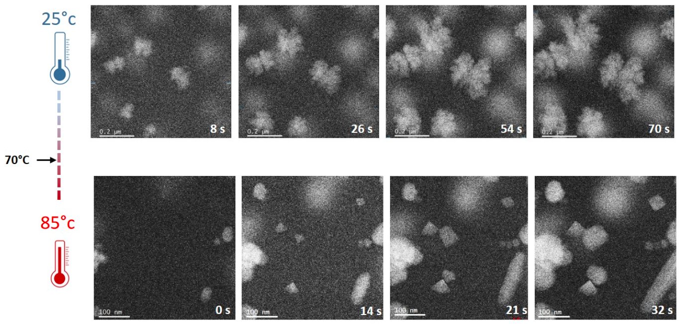
The measurements of nanoparticles growth profiles revealed an increase of the growth speed with the temperature. The evolution of the power law relationship between the mean radius of the nanoparticles and time are compared with growth theory. Beside these kinetic effects, we revealed the drastic impact of the temperature on nanoparticles shape (figure 1): dendritic particles are formed at low temperature (< 70°C) whereas we obtain facetted nanoparticles at high temperature (> 70°C). Surprisingly, this transition in the growth mode of gold nanoparticles is abrupt and occurs at relatively low temperature. All the physico-chemical processes possibly implied in this transition will be carefully discussed.
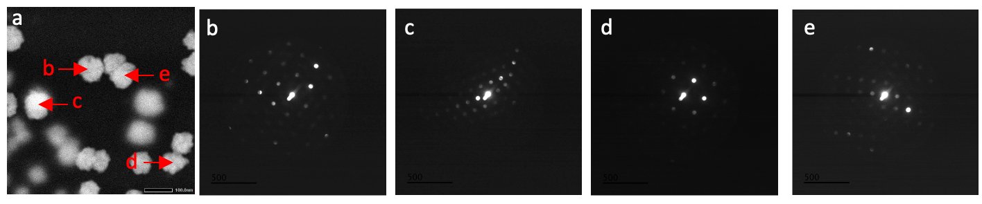
For the first time, we exploited STEM nanodiffraction to study the atomic structure of individual nano-objects during dynamical process in liquid. [4] We will illustrate the advantages of these reciprocal-space investigations with respect to direct imaging. Notably, we will explain the possibility to determine the structure of randomly oriented nano-objects in liquid without spatial resolution limitation and more importantly, the use of very low dose conditions that allows reducing the total dose by almost four order of magnitude with respect to HRTEM analysis. We will show that STEM nanodiffraction can be used during nanoparticles growth experiments, particularly to understand the temperature-induced transition in the growth mechanisms of gold nanoparticles.
Figure 1. In situ STEM HAADF image series of the growth of gold nanoparticles performed at 25°C (top images) and 85°C (bottom images).
Figure 2. (a) In situ STEM HAADF image of the growing gold nanoparticles at 25°C. (b-c-d-e) STEM nanodiffraction. The position of the probe for the acquisition of each diffraction are shown in image (a).
- References
[1] F. M. Ross, Science, Vol. 350 (2015), Issue 6267.
[2] D. Alloyeau et al., Nano Letters, 15 (2015), 5, p.2574-2581.
[3] N. Ahmad et al., Nano Letters, 17 (2017), 7, p. 4194-4201.
[4] A. Khelfa et al., Micron, 116 (2019), p. 30-35.