Thermal Reduction of Transition Metal Oxide Nanocrystals Investigated by in-situ TEM
- Abstract number
- 752
- Event
- European Microscopy Congress 2020
- DOI
- 10.22443/rms.emc2020.752
- Corresponding Email
- [email protected]
- Session
- PSA.5 - Nanoparticles & Catalysts
- Authors
- Xiaodan Chen (2), Heleen van Gog (1), Marijn van Huis (2)
- Affiliations
-
1. Molecular Biophysics, Debye Institute for Nanomaterials Science, Utrecht University
2. Soft Condensed Matter, Debye Institute for Nanomaterials Science, Utrecht University
- Keywords
In-situ TEM, Nanocrystal, Reduction, Transition metal oxide.
- Abstract text
Transition metal oxides (TMOs) display very versatile physical and chemical properties and are consequently used in a wide range of applications, ranging from superconductor to catalysis. Their unique properties stem from their partially filled d orbitals, which allows TMOs to exist in different oxidation states and to exist as various stable polymorphs.[1-3] Understanding of the thermal stablity and their reduction processes is important for controlling their properties in applications. In this study, Co3O4 and TiO2 nanocrystals are investigated as model systems. The reduction of Co3O4 and TiO2 nanocrystals at elevated temperature was investigated by in-situ TEM under vacuum conditions, using a Talos F200X TEM and a DENSsolutions in-situ heating holder.
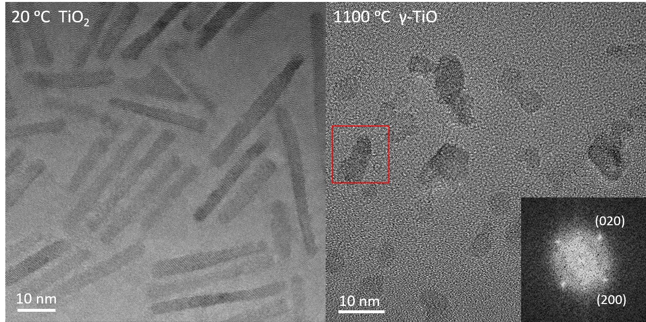
TiO2 anatase nanorods were found to reduce to γ-TiO upon heating to 1100 ⁰C, as shown in Figure 1. Before reduction to TiO, an intermediate phase appeared at 950 ⁰C. From 900 ⁰C onwards, the nanorods broke up into smaller nanocrystal followed by coalescence and crystal fusion of adjacent particles.
Fig. 1 TEM images of anatase TiO2 nanorods at 20 ⁰C before heating (left) and smaller TiO nanocrystals formed after heating to 1100 ⁰C. The inset shows the Fast Fourier Transform (FFT) of the area marked with the red square.
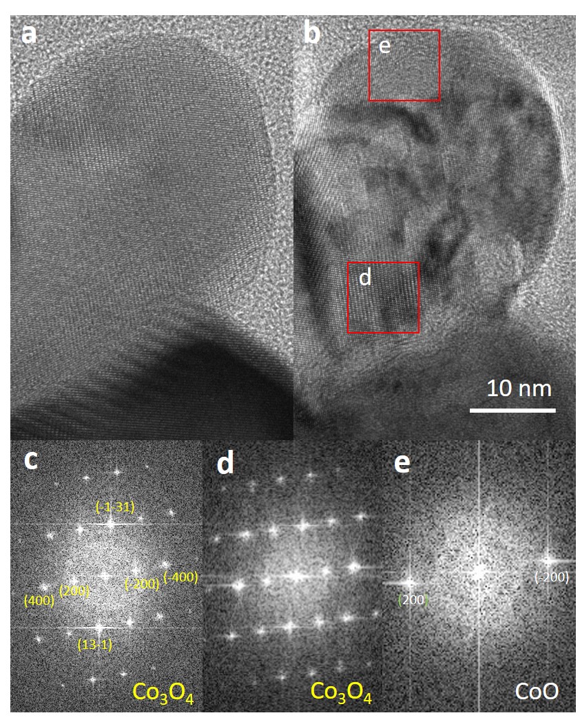
Reduction of spinel Co3O4 nanocrystals to rock salt CoO started at 350 ⁰C and full reduction was obtained after heating to 400 ⁰C for 10 mins.[4] No further reduction was observed when heating to higher temperatures up to 900 ⁰C. The reaction proceeded from the surface to the center of the particle, while the crystal lattices of the parent Co3O4 and the product CoO nanoparticles were in a cube-on cube orientation relationship. The CoO reduced surface layer was sometimes slightly tilted with respect to the Co3O4 interior.
Fig.2 High resolution TEM images of Co3O4 nanocrystals (a) at 20 ⁰C, and (b) at 350 ⁰C; (c) FFT image of (a); (d, e) FFT images of the areas indicated with red squares in (b). Here the surface area was reduced the CoO phase, while the central part is still in Co3O4 phase.
- References
[1] Y. Tokura, N. Nagaosa, Science 2000, 288, 462–468.
[2] P. Poizot, S. Laruelle, S. Grugeon, L. Dupont, J.M. Tarascon, Nature 2000, 407 (6803), 496–499.
[3] H. van Gog, W.F. Li, C. Fang, R.S. Koster, M. Dijkstra, M. van Huis, npj 2D Mater. Appl. 2019, 3 (1), 1–12.
[4] X.D. Chen, H. van Gog, G.A. Blab, M.A. van Huis, in preparation.