WHOLE ORGAN QUANTITATIVE BIOLOGY WITH LIGHT SHEET MICROSCOPY
- Abstract number
- 57
- Event
- European Microscopy Congress 2020 Invited Speakers
- DOI
- 10.22443/rms.emc2020.57
- Corresponding Email
- [email protected]
- Session
- LSA.2 - Dynamic interactions in cells, organoids, tissue and entire organisms
- Authors
- Prof. Dr. Matthias Gunzer (1)
- Affiliations
-
1. Institute for Experimental Immunology and Imaging, University Hospital, University Duisburg–Essen
- Keywords
bone vascular system
ischemia/reperfusion injury
kidney vascular system
Light sheet microscopy
tissue clearing
- Abstract text
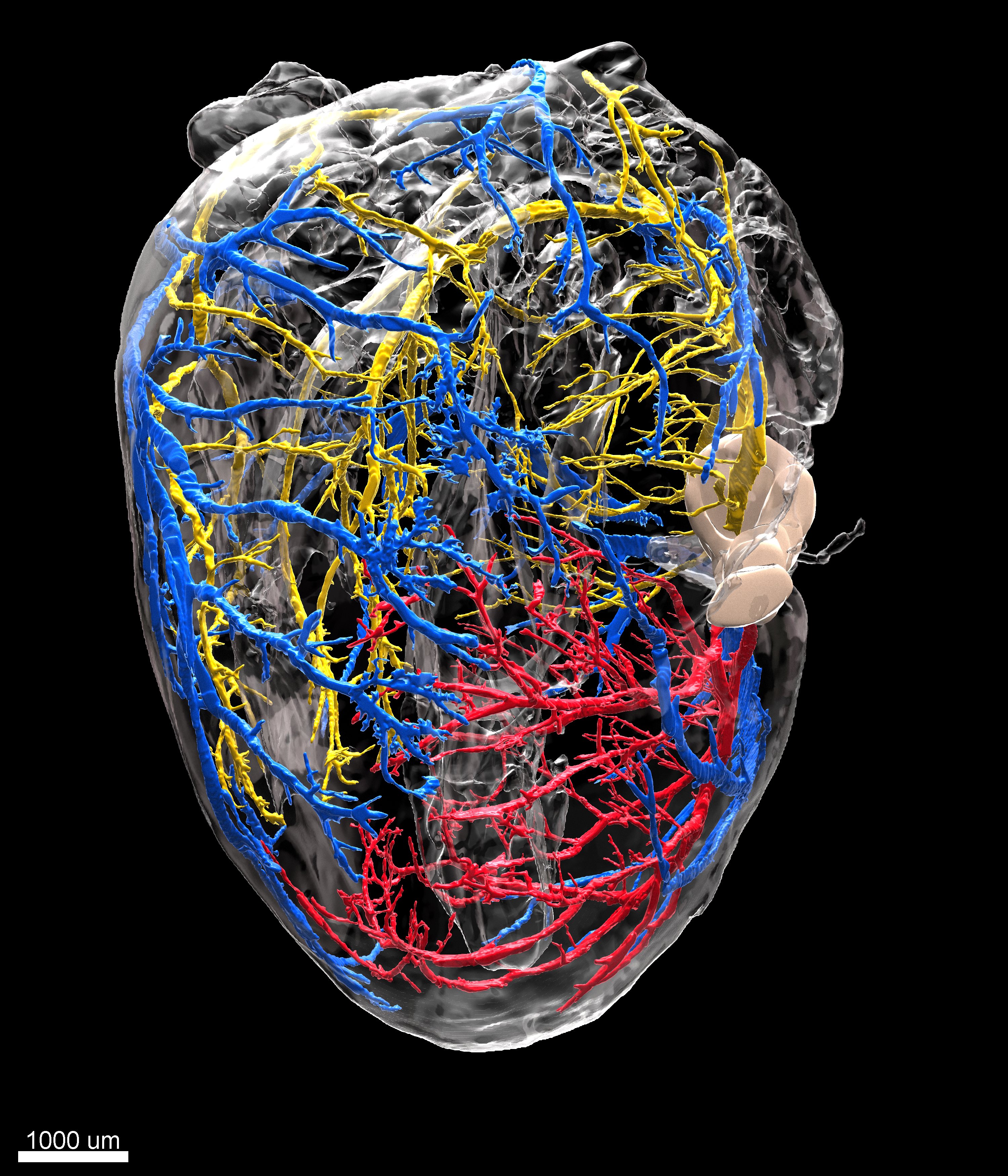
Figure 1: LSFM of the blood vessel system in a murine heart suffering from I/R injury. Red are the vessels, which are occluded by the knot visible in grey on the right of the heart. Major arteries are in yellow, major veins in blue. They grey channel shows autofluorescence to demonstrate the overall organ structure.
Tissue clearing (TC) and imaging by light sheet fluorescence microscopy (LSFM) has become a powerful tool to study the overall composition of functional units in entire organs as well as the impact of diseases on these structures. I will show how we have made use of TC/LSFM to investigate the blood vessel system and immune infiltrate of murine organs. First, we have investigated the blood supply of long bones, thereby discovering and functionally characterizing an entirely new blood vessel system that is responsible for the great majority of blood and leukocyte transport into and out of bones. Furthermore, we could show how ischemia/reperfusion injury has an impact on the blood vessel system of mouse hearts and how neutrophil granulocytes infiltrate the organ at disease-relevant sites. Finally, we were able to obtain a whole organ view on functional units (glomeruli) of murine kidneys, that allowed to precisely quantify these structures with automated image analysis. The impact of autoimmune disease was then very obvious as it massively influenced both, the number and size of glomeruli. Hence, TC/LSFM is extremely useful to get a mesoscopic, whole organ view on normal and pathological physiology including precise quantitative data on functional elements responsible for organ operation.
- References
1. Merz SF, Korste S, Bornemann L, Michel L, Stock P, Squire A, Soun C, Engel DR, Detzer J, Lörchner H, Hermann DM, Kamler M, Klode J, Hendgen-Cotta U, Rassaf T, Gunzer M, Totzeck M. Contemporaneous 3D characterization of acute and chronic myocardial I/R injury and response. Nat Commun. 2019;10(1):2312.
2. Gruneboom A, Kling L, Christiansen S, Mill L, Maier A, Engelke K, Quick HH, Schett G, Gunzer M. Next-generation imaging of the skeletal system and its blood supply. Nat Rev Rheumatol. 2019;15(9):533-549.
3. Grüneboom A, Hawwari I, Weidner D, et al. A network of trans-cortical capillaries as mainstay for blood circulation in long bones. Nat Metabolism. 2019;1(2):236-250.
4. Klingberg A, Hasenberg A, Ludwig-Portugall I, Medyukhina A, Männ L, Brenzel A, Engel DR, Figge MT, Kurts C, Gunzer M. Fully Automated Evaluation of Total Glomerular Number and Capillary Tuft Size in Nephritic Kidneys Using Lightsheet Microscopy. J Am Soc Nephrol. 2017;28(2):452-459.