Workflow for correlative energy-dispersive X-ray tomography and atom probe tomography
- Abstract number
- 327
- Event
- European Microscopy Congress 2020 Invited Speakers
- DOI
- 10.22443/rms.emc2020.327
- Corresponding Email
- [email protected]
- Session
- DHA.3 - Machine assisted acquisition and analysis of microscopy data
- Authors
- Dr Amandine Verguet (1), Mr Martin Jacob (4), Dr Jyh-Miin Lin (4), Dr Philippe Ciuciu (2), Dr Pascale Bayle-Guillemaud (3), Dr Isabelle Mouton (1), Dr Zineb SAGHI (4)
- Affiliations
-
1. CEA, DEN, Service de Recherches Métallurgiques Appliquées (SRMA), Université Paris-Saclay, F-91191
2. CEA/DRF/Joliot NeuroSpin, Université Paris-Saclay, F-91191
3. Univ. Grenoble Alpes, CEA-IRIG, MEM, F-38000
4. Univ. Grenoble Alpes, CEA, LETI, F-38000
- Keywords
APT, correlative tomography, EDX, electron tomography, open-source packages
- Abstract text
The past decade has seen the development of correlative tomography for the extraction of 3D complementary information about the same sample, at different scales and resolutions [1]. This approach has brought new insights into the structural and chemical properties of materials. However, numerous challenges have emerged concerning big data handling, hyperspectral image analysis and volume registration that takes artefacts inherent to each tomography modality into account. In this work, we combine high angle annular dark field (HAADF)-STEM tomography, energy-dispersive X-ray (EDX) tomography and atom probe tomography (APT) to investigate the size, composition and morphology of nano-precipitates in an oxide dispersion-strengthened (ODS) steel [2]. We present a correlative analysis workflow that aims at overcoming the limits imposed by each technique alone.
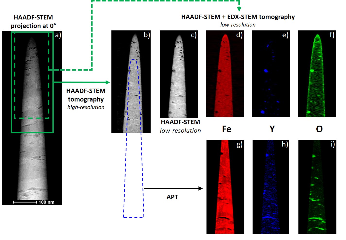
A needle-shaped sample of the ODS material, composed of an Fe-Cr matrix and yttrium aluminium titanium oxide nano-precipitates, is prepared by focused ion beam (FIB). Figure 1(a) shows an HAADF-STEM image of the needle. A first region is selected for the HAADF-STEM tomography experiment (green rectangle). The standard simultaneous iterative reconstruction technique (SIRT) is used for the 3D tomographic reconstruction. An orthoslice through the volume (Figure 1(b)) shows well-resolved block-shaped and spherical nano-precipitates, but does not provide information about their composition. A sub-region (dashed green rectangle in Figure 1(a)) is chosen for the 3D chemical analysis of the nano-precipitates by EDX-STEM tomography. Hyperspectal data analysis is performed using Hyperspy library, and a compressed sensing algorithm based on high-order total variation (HOTV) and implemented in the open-source PySAP library [3,4] is employed for the 3D reconstruction of elemental volumes. Figure 1(d,e,f) are orthoslices through the Fe, Y and O volumes. An in-house code for volume registration is applied to correlate the highly resolved HAADF-STEM volume (~1nm resolution) and the chemically sensitive EDX-STEM volumes (~5nm resolution). The sample is then transferred to be analysed by APT. The dashed blue line in Figure 1(b) indicates the position of the reconstructed volume compared to the electron tomography experiments. Information about the position and chemical composition of nano-precipitates is retrieved at the sub-nanometer scale, with uncertainties related to the difference in field evaporation between the nano-precipitates and the matrix. Fe, Y, and O orthoslices through the reconstructed volumes are shown in Figure 1(g,h,i). Distortions in the APT volumes are identified based on the HAADF-STEM and EDX-STEM reconstructions. For this purpose, a python-based package is developed with non-rigid registration tools and an iterative optimization approach for the choice of APT reconstruction parameters.
We will show that the improved APT volumes provide information about nano-precipitates that electron tomography cannot resolve due to limited composition sensitivity or spatial resolution [5].
Figure 1. Correlative HAADF-STEM/EDX-STEM/APT workflow. (a) HAADF-STEM image of the ODS sample. (b) Orthoslice through the high-resolution HAADF-STEM reconstruction (corresponding to the green rectangle in (a)). (c) Orthoslice through the low-resolution HAADF-STEM reconstruction (corresponding to the dashed green rectangle in (a)). This same region is selected for the EDX-STEM tomography, and the volume analyzed by APT is indicated in dashed blue in (b). (d,e,f) and (g,h,i) are orthoslices through the Fe, Y and O of the EDX-STEM and APT reconstructions, respectively.
- References
[1] T.L. Burnett and P.J. Withers, Nature Materials 2019, 18: 1041.
[2] C. Capdevilla, M.K. Miller et al., Materials Science and Engineering 2008, 490: 277.
[3] M. Jacob, J.M. Lin et al., EMC Copenhagen 2020.
[4] S. Farrens, A. Grigis et al., Astronomy and Computing 2020, in revision.
[5] This work was carried out at the Nanocharacterization Platform (PFNC) of Minatec, supported by the ‘Recherche technologique de Base’ program of the French ministry of Research. We acknowledge the financial support of the Cross-Disciplinary Program on Numerical Simulation of CEA, the French Alternative Energies and Atomic Energy Commission. This work profited from a French government grant managed by the National Agency of Research under the program ‘Investments for the future’ (ref. ANR-11-EQPX-0020).