X-EDS electron tomography and clustering in EELS applied to the resolution of rare earth doped CeO2 mesoporous structures.
- Abstract number
- 959
- Event
- European Microscopy Congress 2020
- DOI
- 10.22443/rms.emc2020.959
- Corresponding Email
- [email protected]
- Session
- DHA.2 - Advances in 3-dimensional image reconstruction
- Authors
- Javier Blanco-Portals (3, 5), Dr Federico Biautti (1), Simone Anelli (1), Dr Marc Torrell (1), Dr Miguel López-Haro (2), Dr Sònia Estradé (3, 5), Dr Albert Tarancón (1, 4), Professor Jose Calvino (2), Professor Francesca Peiró (3, 5)
- Affiliations
-
1. Catalonia Institute for Energy Research (IREC), Department of Advanced Materials for Energy, 1 Jardins de les Dones de Negre,08930
2. Dep. de Ciencia de los Materiales e Ingeniería Metalúrgica y Química Inorgánica Facultad Ciencias Universidad de Cádiz. Campus Rio San Pedro, Puerto Real, 11510
3. LENS-MIND, Departament d'Enginyeria Electrònica i Biomèdica, Universitat de Barcelona, 08028
4. ICREA, 23 Passeig Lluís Companys, 08010
5. Institute of Nanoscience and Nanotechnology (IN2UB), Universitat de Barcelona, 08028
- Keywords
Cerium Oxide, EDS, EELS, Electron tomography, Mesoporous.
- Abstract text
Cerium Oxide nanostructured materials have remained a hot topic in materials science in recent years. CeO2 mesoporous structures have been rutinarly proposed as scaffold structures for the electrodes in solid state oxide fuel cells (SOFC) and solid oxide electrolyser cells (SOEC). CeO2 is by nature a good O-ion conductor, that can be easaly synthetised as a mesoporous material giving large surface areas and high porosity, and impregnated by an electronic conducting materials (e.g. transition metals perovskites), providing nanocomposite with mixed ionic and electronic conducting (MIEC) behaviour [1].
Here, we present the characterization of a mesoporous nanostructured CeO2 doped with Gd and Pr. The presence of these dopants is known to allow for MIEC behaviour without the inclusion of an electron conducting material interface [2], [3]. Variations of the electrical conductivity across integrated devices with different dopant ratios have been measured, showing a clear dependency on the dopant nature and overall stoichiometry. The hypothesis is that the conductivity in such mesoporous CeO2 materials is bounded to the distribution of dopants and possible preferencial segregation towards grain boundaries and grain surfaces and, thus, a precise analytical characterization of the spatial morphology becomes mandatory for a fine control of the devices performance.
Due to the high complexity of the interconnected double-gyroid mesoporous morphology, only small localized regions of grain boundaries in the outer corona of the mesoporous strucure could be analyzed by planar EELS to avoid the severe overlapping of signals in thicker areas. Clear segregation of dopants towards the grain boundaries was resolved applying the K-means clustering analysis[4]. Nevertheless, the spatial distribution of dopants across the whole structure (inner regions of the mesoporous) remained unresolved.
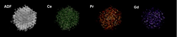
To address this issue, we undertook an analytical electron tomography experiment centred on the X-EDS signals, making the most of the capabilities of the TVM3D algorithm, achieving succesful results under severe undersampling conditions (9 projections, from -70º to +70º each 20º and including the 0º projection)[5]. Under these kind of acquisition conditions, classic algorithms such as SIRT would have failed.
Dopant segregation was confirmed throughout the entire structure, showing the higher concentration regions located in the grain intersection areas (boundaries) and surfaces.
Figure 1. Volume rendering of the reconstructions for the (from left to right) ADF signal and the X-EDS Ce, Pr and Gd signals.
Acknowledgements
This work has been supported by the Spanish Ministerio de Economía y Competitividad through the projects MAT2016-79455-P and RED IMAGINE MAT2016-81720-REDC, and the government of the Generalitat de Catalunya (FI grant 2018FI_B_00360). This work has also been possible thanks to the collaboration with Dr.Emerson Coy and Professor Stefan Jurga from the Adam Mickiewicz University (Poznan, Poland).
- References
[1] E. Hernández, F. Baiutti, A. Morata, M. Torrell, and A. Tarancón, “Infiltrated mesoporous oxygen electrodes for high temperature co-electrolysis of H2O and CO2 in solid oxide electrolysis cells,” J. Mater. Chem. A, vol. 6, no. 20, pp. 9699–9707, 2018.
[2] L. Almar et al., “A Durable Electrode for Solid Oxide Cells: Mesoporous Ce0.8Sm0.2O1.9 Scaffolds Infiltrated with a Sm0.5Sr0.5CoO3-δ Catalyst,” Electrochim. Acta, vol. 235, pp. 646–653, 2017.
[3] M. Acosta, F. Baiutti, A. Tarancón, and J. L. MacManus-Driscoll, “Nanostructured Materials and Interfaces for Advanced Ionic Electronic Conducting Oxides,” Adv. Mater. Interfaces, vol. 6, no. 15, pp. 1–15, 2019.
[4] P. Torruella et al., “Clustering analysis strategies for electron energy loss spectroscopy (EELS),” Ultramicroscopy, vol. 185, pp. 42–48, Feb. 2018.
[5] M. López-Haro et al., “A Macroscopically Relevant 3D-Metrology Approach for Nanocatalysis Research,” Part. Part. Syst. Charact., vol. 35, no. 3, p. 1700343, Mar. 2018.