XFold slides for fluorescence microscopy imaging
- Abstract number
- 791
- Event
- European Microscopy Congress 2020
- DOI
- 10.22443/rms.emc2020.791
- Corresponding Email
- [email protected]
- Session
- LSA.3 - Applications for imaging sub-cellular events at high resolution
- Authors
- Dr Nagarajan Subramaniyam (2), Mr Jonas Ylönen (2), Dr Antti Isomäki (1)
- Affiliations
-
1. University of Helsinki
2. Xfold Imaging Oy
- Keywords
Fluorescence microscopy, photobleaching, hot spots, focal adhesion
- Abstract text
Fluorescence microscopy is a widely used method for imaging various biological samples, which relies on using fluorescent dyes to image areas of interest. Despite the wide range of fluorescent dyes available, some imaging applications suffer from a low fluorescence signal from the dyes used in them. All fluorescent dyes also suffer from rapid photobleaching when excited with high laser powers, preventing long imaging times of one area, or alternatively requiring a low power to be used, which leads to poor image quality. Different types of patterned metal surfaces and metallic nanoparticles have been demonstrated to enhance the fluorescence signal, but these enhancements have typically not been significant and are concentrated on “hot spots” around the metal features.
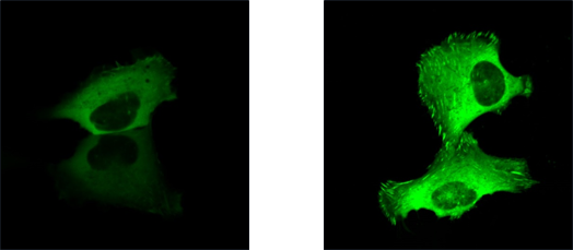
Xfold slides are designed to enhance the sensitivity of fluorescence microscopy in a close range from the slide surface, in order to better visualize the focal adhesions. Xfold slides provide a significantly enhanced fluorescence signal, allowing a lower laser power to be used in imaging, and enabling improved imaging in cases where the fluorescence signal is typically low. The slides work uniformly over the entire surface area, without large variations in enhancement or “hot spots”. Xfold slides also improve the cell adhesion to the surface.Figure 1: U2OS cells with Talin GFP imaged with 40% laser power on a glass slide (left), and 2% laser power on Xfold slide (right).